JOURNAL OF THE
ROYAL MEDICAL SERVICES

Official Publication for the Jordanian Royal Medical Services


MODIFIED MITCHELL’S OSTEOTOMY FOR THE TREATMENT OF HALLUX VALGUS. EXPERIENCE AT KING HUSSEIN MEDICAL CENTER


Mohamed Zaher, MD*, Issam Dahabra, MD*


ABSTRACT

Objective: To evaluate the outcome of modified Mitchell’s osteotomy for treatment of hallux valgus.

Methods: We reviewed the results of 69 patients (84 feet) in whom the modified Mitchell’s osteotomy with Y shape advancement of medial capsule, lateral adductor tenotomy and lateral capsule release, was done between 1993-2001 with an average follow up of 30 months (range 10-48 months).

Results: Radiographic and subjective results were evaluated. Preoperative criteria included intermetatarsal angle of greater than 14 degrees (average 16.16), average metatarsophalangeal angle was 38.4 degrees (range 32-48 degrees) and painful Hallux valgus deformity. The average postoperative intermetatarsal angle was 7.9 with relative correction of 8 degrees. The average postoperative metatarsophalangeal angle was 9.2 degrees (range from 2-18). Subjectively, all patients were satisfied with their results and stated they would have the procedure again. There were no complications noted in the patients such as avascular necrosis, Hallux varus, and recurrence of deformity. There were 6 delayed union cases for 6 months, and one infection.

Conclusion: The modified Mitchell’s osteotomy is an appropriate operative method that produces satisfactory results, and can be recommended in the treatment of moderate to severe Hallux valgus.

Key words: Modified Mitchell’s Osteotomy, Hallux valgus, Surgical outcome.

JRMS Dec 2003; 10(2): 6-10


Introduction

Hallux valgus is a complex deformity of multifactorial etiology that causes pain, different shoe fitting, and dissatisfaction with the appearance of the foot. There are over 130 surgical procedures for the treatment of Hallux valgus, some include soft tissue procedures, others bony procedures and some combine both soft tissue and bony correction.

Hallux valgus was an uncommon deformity of the foot, until wearing fully enclosed shoes and boots was introduced (1-7).

The earliest mentioning of such deformity in the literature was in the eighteenth century. It is rarely seen in those who do not wear shoes.
Hawkins and associates (1) the first described the Mitchell operation for hallux valgus in literature in 1945.

C. Leslie Mitchell subsequently published an article in 1958 describing this procedure, and from this point on it became known as Mitchell bunionectomy including osteotomy of the distal portion of the first metatarsal, lateral displacement and angulation of the head of the metatarsal and bunionectomy and capsulorrhaphy (1,2,8,9).

The modified Mitchell’s osteotomy with Y-shape advancement of the medial capsule and lateral adductor tenotomy is recommended for correction of moderate to severe hallux valgus when pain and discomfort persist despite conservative treatment (1-3,8,10).

Numerous surgical procedures for correction of this deformity have been described, and the choice of procedure depends primarily on severity, location, and nature of the pathology.

Many factors are considered for Mitchell’s osteotomy, including general health, level of pain, range of movement, quality of bone and tissue, life style, foot wear and age (4,11-13).

The indications for Mitchell’s hallux valgus corrective osteotomy include:
1.    Young patients with severe hallux valgus.
2.    Moderate to severe pain
3.    Deformity and joint instability.
4.    Inability to wear normal footwear.
5.    Failure of conservative treatment to restore the foot problem.

This procedure has been used at King Hussein Medical Center since 1993 for the correction of moderate and severe hallux valgus deformity of intermetatarsal (IM) angle >14º, and metatarsophalangeal (MTP) angle >32º. The purpose of this retrospective study was to review the results of all patients who have undergone modified Mitchell’s osteotomies with Y-shape advancement of medial capsule and lateral adductor tenotomy for hallux valgus between 1993 and 2001.

The IM angle is formed by the intersection of longitudinal axes of the first and second metatarsals. A normal angle is less than 9 degrees. The metatarsophalangeal angle (MTP) is formed by the intersection of longitudinal axes of the first metatarsal and the proximal phalanx. A normal angle is less than 15 degrees. (Fig. 1).

 m.zaher1.PNG
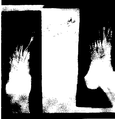
Fig. 1:
Intermetatarsal (IM) angle and the metatarsophalangeal angle (MTP)


Methods

We retrospectively studied 69 patients (84 feet) in whom modified Mitchell’s osteotomies with Y-shape advancement of the medial capsule and lateral adductor tenotomy were performed between 1993 and 2001 at King Hussein Medical Center in Jordan, for moderate to severe and painful hallu valgus, with average follow up of 30 months (10-48 months).

Female patients were 51; male patients were 18 (female to male ratio 2.8:1) right side 38 feet, left side 31 feet, and bilateral 30 feet in 15 patients.
The patients ranged in age from 16-47 years (average 23.7 years).

Indications for surgery included shoe problems, persistent pain and discomfort despite conservative management. Two surgeons performed the operations.

Weight-bearing dorsoplanter and lateral radiographs of the foot were obtained preoperatively and postoperatively and IM and MTP angles were measured. (Figs. 2, 3).


m.zaher2.PNG
Fig.  2:  Preoperative weight bearing radiographs.
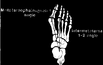

 m.zaher3.PNG
Fig. 3: Postoperative weight bearing radiographs.


The Modified Mitchell is Osteotomy
The Mitchell is osteotomy is a metatarsal osteotomy that displaces the metatarsal head slightly lateral ward in order to correct the hallux valgus deformity. This procedure can be conducted in deformities of any degree of severity.

 Surgical Technique
Longitudinal skin incision was centered over the dorsomedial aspect of the first metatarso-phalangeal joint and extended from the mid-portion of the proximal phalanx to the mid portion of the first metatarsal.

A V-Y capsular flap was created over the medial aspect of the MTP joint.
The medial eminence was exposed and the osteotomy created through a transverse cut in the metatarsal neck leaving a lateral piece of cortex about 3 mm wide. The second cut was made 2 mm more proximal and parallel to the first cut in order to create a step-off in the distal fragment.

Two dorsal to planter drill-holes were made: One in the distal fragment and the second hole in the proximal fragment about 1 cm apart.

The capital fragment was then displaced laterally until the lateral shelf locked over the lateral aspect at proximal fragment of the shaft.

The site of osteotomy was then fixed using non-absorbable sutures, through the two dorsal to planter drill-holes.

Capsular closure (medial capsulorrhaphy) The V-shaped incision then advanced into the straight limb of the Y-shaped incision while holding the hallux in planter flexion.

A 3 cm dorsal longitudinal incision was made centered over the first intermetatarsal space to expose the conjoined adductor tendon, the tendon was dissected from the lateral sesamoid bone, and the transverse intermetatarsal ligament was released. (Fig. 4).


m.zaher4.PNG
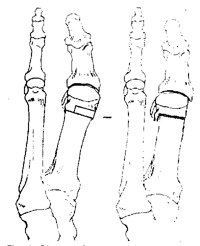
Fig. 4:
Diagrammatic representation of Mitchell’s osteotomy.


Post-Operative Care
The patients remained non-weight bearing with crutches until sutures were removed at 14 days post operation. The foot was then placed in a non weight-bearing fiber- plaster cast which was removed at 6-8 weeks post operatively, after which walking was permitted with the foot immobilized in a below-knee cast until the site of osteotomy healed at 10 to 12 weeks, post-operatively.


Results

The patients were assessed before undergoing the operation and at follow-up of average of 30 months after operation. Assessment was made by interviews and clinical examination by the two authors. (Table I)

  Table I:Clinical results in 69 patients.

Results

Satisfied

No. of patients

Satisfied with reservation

No. of patients

Dissatisfied

No. of patients

Pain

65

4

0

Appearance

67

2

0


It was found that patients with an IM angle <15º were absolutely pain-free and satisfied with the outcome of the operation.

Eighty six percent of patients had no shoewear problems, 14% had minor difficulty with shoe width or heel height, especially old aged women. No patients had to wear orthotic shoes.  Subjectively, 92% of the patients rated their satisfaction with the outcome of the operation as excellent or good, 8% were satisfied with the outcome of the procedure. 

 Table II:  Functional Grading (Adapted from the assessment proposed by the American Orthopedic Foot and Ankle Society).

Grade

Pain

Pre Op.

Post Op.

0

None


1

During sports & other vigorous activities

45

0

2

With conventional shoes in daily walking

84

0

3

Walking bare footed

15

0

4

At rest

10

0


The average preoperative and IM angle was 16.2 in the cases studied, the average postoperative IM angle was 7.9 with a relative correction of 8 degrees. (Table III).

  Table III: Pre-operative and post-operative IM angles.

No. of Cases

Pre-operative

No. of Cases

Post-operative


IM angle


IM angle

15

14

19

5

24

15

20

6

12

16

9

7

12

17

16

8

8

18

11

9


The average preoperative MTP was 38.4 degrees (range 32-48 degrees), the average postoperative MTP angle was 9.2 degrees (range from 2-18 degrees). (Table IV).

 Table IV:Preoperative and postoperative MTP angles

No. of Cases

Pre-operative

No. of Case

Post-operative


MTP angle


MTP angle

9

32

8

2

12

34

11

4

12

35

18

6

8

36

16

10

8

38

10

12

11

40

8

14

5

42

9

16

10

44

4

18

9

48


84

Average 38.4

84

Average 9.2


Complications
The two main complications were delayed union in 6 feet (7.1%) of the cases and infection in one foot (1.2%) of the cases.

Only 6 feet had delayed union for 6 months, and all other feet were united within 12 weeks.

The infection which occurred in one patient, was treated by administration of antibiotics and daily dressing. At the latest follow-up examination, the patient experienced no pain, had no restriction concerning shoewear, and was satisfied with the outcome of the operation.


Discussion

Numerous procedures have been described for the correction of hallux valgus deformity (1,2,4,6,10-14). Most authors agree that severe hallux valgus deformity with marked metatarsus primus varus (IM angle >15º) can be surgically corrected by distal osteotomy (8,9,11,15).

The Mitchell procedure can produce satisfactory realignment of the first metatarsophalangeal joint (1-3,8).

This is the first study conducted at King Hussein Medical Center focusing on moderate to severe hallux valgus. Our series with mean preoperative values for IM angle 16.2 confirms the potential of the Modified Mitchell’s Osteotomy to correct even severe cases of hallux valgus and metatarsus primus varus. Mean correction for IM angles (9.7) proved to be sufficient in the majority of cases in correcting the pathology to normal values to a mild residual deformity.

All our osteotomies have united despite 6 cases of delayed union. No patient had serious complication; the patient who developed superficial infection was due to tight closure of the skin and superficial necrosis. No patient required orthotic shoewear after the operation. No patient was dissatisfied with the outcome. The incidence of varus and recurrent valgus deformity had not increased from the initial follow-up study.

Comparison with other operations for hallux valgus is difficult since so many different protocols have been used for assessment (9,12,13,15-17). We have adopted the American Orthopedic Foot and Ankle Society (Smith et al 1984) (18). Our results in terms of correction of valgus angle, IM angle, and range of movement of the toe compared favorably with those reported by Mitchell et al (1958) (2).


Conclusion

The modified Mitchell is osteotomy is an appropriate and effective procedure in correcting hallux valgus deformity with IM angle >14º and should be considered in the surgical management of patients with moderate to severe symptomatic hallux valgus associated with metatarsus primus varus.


References

1. Hawkins F, Mitchell C, Hedrick D.  Correction of hallux valgus by metatarsal osteotomy. J Bone Joint Surg 1945; 27: 387-398.

2.  Mitchell CL, Fleming J, Allen R, et al.  Osteotomy-bunionectomy for hallux valgus. J Bone Joint Surgery 1958; 40(A): 4.

3.  Canale PB, Aronsson DD, Lamont Rl, et al.  The Mitchell procedure for the treatment of adolescent hallux valgus. The Journal of Bone and Joint Surgery 1993; 75-A: (11): 1610-1618.

4. Torrkki M, Malmivaara A, Seitsalo S, et al. Surgery vs orthosis vs watchful waiting for hallux valgus. JAMA  2001; 5(12): 20-26.

5.  Saltzman Cl, Aper RL, Brown TD. Anatomical determinants of first metatarsophalangeal flexion moments in hallux valgus. Clinical Orthopaedics and Related Research 1997; 339(6): 261-269.

6. Clayton ML, Leidholt JD, Clark W. Arthroplasty of rheumatoid metatarsophalngeal joints. Clinical Orthopaedics and Related Research 1997; 340(7): 48-57.

7. Kitaoka HB, Platzer Gl.  Salvage treatment of failed hallux valgus operations with proximal first metatarsal osteotomy and distal sort-tissue reconstruction. Foot Ankle Int 1998; 19(3): 127-131.

8.  Briggs TWR, Smith P, Mcauliffe TB. Mitchell’s osteotomy using internal fixation and early mobilization. The Journal of Bone and Joint Surgery 1992; 74-B:(1): 137-139.

9. Skalley TC, Myerson MS. The operative treatment of acquired hallux varus. Clinical Orthopaedics and Related Research 1994; 306: 183-191.

10. Sangeorzan BJ, Hansen ST. Modified Lapidus Procedure for Hallux Valgus. Foot & Annkle 1989; 9(6): 262-266.

11.  Resch S, Stenstrom A. Proximal closing wedge osteotomy and adductor tenotomy for treatment of hallux valgus. Foot & Ankle 1989; 9(6): 272-280.

12. Jones KJ, Feiwell LA, Freedman EL, et al. The effect of chevron osteotomy with lateral capsular release on the blood supply to the first metatarsal head. The Journal of Bone and Joint Surgery  1995; 77(2): 197-204.

13.  Resch S, Stenstrom A, Reynisson K, Jonsson K. Chevron osteotomy for hallux valgus not improved by additional adductor tenotomy. Acta Orthop Scand 1994; 65(5): 541-544.

14.   Mann RA, Rudicel S, Graves SC. Repair of hallux valgus with a distal soft tissue procedure and proximal metatarsal osteotomy. The Journal of Bone and Joint Surgery 1992; 11: 124-132.

15.  Trnka Hj, Kabon B, Zettl R, et al. Helal metatarsal osteotomy for the treatment of metatarsalgia: A critical analysis of results. Acta Orthop Scand 1996; 19(5): 457-461.

16. O’Malley MJ, Chao W, Thompson FM. Treatment of established nonunions of Mitchell osteotomies. Foot Ankle Int 1997; 18(2): 77-80.

17.   Laughlin TJ. Complications of distal first metatarsal osteotomies. J Foot Ankle Surg 1995; 34(6): 524-531.

18.  Smith RW, Reynolds JC, Stewart MJ. Hallux valgus assessment: Report of Research Committee of the American Orthopedic Foot and Ankle Society. Foot Ankle 1984; 5(5): 92-103.


About
The Journal

The Journal of the Royal Medical Services (JRMS) is an open access journal and it is the official publication for the Royal Medical Services of the Jordanian Armed Forces... Read More

Subscribe to OUR
newsletter

To receive updates on new issues

JRMS Journal

Articles Archive

Archive

Previous Issues

Volume 25
April 2018

Volume 24
December 2017

Volume 24
August 2017

Volume 24
March 2017