ABSTRACT
Objective: To study the structural response
of the palatal epithelium to the fitting of new complete maxillary
acrylic dentures in a group of subjects over a six-month period.
Methods: Twenty-five
edentulous subjects, 13 females, and 12 males had new acrylic dentures
fabricated by standard procedures in the prosthetic department of the
Institute of Dental Surgery, London. Exfoliative cytology was used to
collect surface cells from the lateral wall of the palate in the region
of the second premolar. Pressure sensitive tape was employed to sample
each subject at insertion of the dentures and after periods of one week
and one, three and six months. Each sample was fixed in 95% ethyl
alcohol, stained, and examined in a light microscope. About 200-350
epithelial cells were counted in each sample and the numbers of fully
keratinized, non-nucleated cells were recorded to calculate the degree
of keratinization. Statistical analysis was performed using the
Mann-Whitney U test.
Results: A significant difference
(p<0.05) in keratinization levels was found between the male and
female subjects. The mean percentage keratinization at insertion was
41.9% for males and 81% for females. After six months of wearing the new
dentures the mean percentage keratinization was 30.4% for males and
87.8% for females.
Conclusion: The wearing of complete
maxillary acrylic dentures disturbs the denture supporting epithelium of
the palatal mucosa. The mean levels of keratinization of male and
female subjects vary significantly, and showed periodic increases and
decreases in individual subjects over a six-month period.
Key words: Keratinization, Complete denture, Cytology
JRMS June 2005; 12(1): 25-29
Introduction
The effect of dentures upon the oral mucosal membrane has been the subject of a considerable number of research investigations. The results of many of these are contradictory; some studies suggested that the denture bearing epithelium become more keratinized while others found that the tissues become parakeratinzed (1-2). The techniques used to sample the tissues in such studies may account for the confusion of the findings. Studies, which have examined histological preparations of biopsies material, had been limited by the difficulty of performing biopsies in large number of patients and the dissimilarity of tissue sites selected for biopsy.
Exfoliative cytology has proved more appropriate for examining the keratinization of the oral mucosa
especially the changes seen in response to denture wearing.
Traditionally, the cytological sampling of the mucosa has been performed using a wooden tongue blade (3). However, another technique using adhesive tape to collect the superficial epithelial squames was proposed by Harinasuta (4) and used to examine levels of palatal keratinization in fully dentate subjects and in subjects wearing tooth supported cobalt-chromium partial dentures. The study confirmed that the adhesive tape technique accurately determines the levels of keratinization of the tissues.
This investigation was designed to evaluate the structural response of palatal epithelium to the fitting of new complete maxillary acrylic dentures in a group of subjects over a six-month period.
Methods
A group of edentulous patients programmed to have new dentures made by the postgraduate students of the Department of Prosthetic Dentistry, in the Institute of Dental Surgery, London was chosen for the study. They were 25 edentulous patients, 13 females and 12 males who were undergoing treatment for the provision of new dentures, due to their previous dentures being ill fitting. Complete dentures were fabricated according to the standard principles followed in the department. The teeth used were Myersons Dura-Blend (Myerson Tooth Corporation, Cambridge, MAAS, USA) using 30o cusp angles; the occlusal scheme provided adequate free way space, intercuspal position coincident with the retruded contact position, and balancing occlusal contacts in lateral and protrusive excursions. Each subject was assessed clinically for the presence or absence of inflammation of the palatal mucosa, as indicated by the presence of erythema and possible petechiae. The age, sex, smoking habits, and general health history of each subject were recorded.
The degree of keratinization of the denture-bearing epithelium of the maxilla was investigated by cytological sampling according to the method of Harinasuta (4).
Each patient had samples taken at the following times:
1. Prior to insertion of complete denture (old denture wearing).
2. One week after insertion.
3. One month after insertion.
4. Three months after insertion.
5. Six months after insertion.
The sites for sampling were chosen in the region halfway between the gingival margin and the midline of the palate at a level corresponding to the 1st premolar. The subjects did not rinse their mouths before the samples were taken. The adhesive tape used was 3M Pressure Sensitive Tape (3M Ltd., Bracknell, Berkshire). The area of the palate was first dried by air syringe, a strip of adhesive tape sized 2cm x 1.5cm was then attached to the palate, using sufficient pressure in order to adhere to the tissue. Then the strip of adhesive tape was peeled off and immediately placed in 95% ethyl alcohol for about 15 minutes. It was then transferred from 95% ethyl alcohol and placed on a clear glass slab with the sticky side facing upward until it was completely dry.
During the drying period the glass slab was covered in order to protect the strip of adhesive tape from dust. Then the strip of adhesive tape was mounted on a clear glass slide with DPX mounting solution (BDH Chemicals Ltd., Poole, Dorset). The slide was labeled and dried completely, ready for staining.
The adhesive tape samples were fixed and stained with Papanicolaou’s (5) stain and examined by light microscopy. The stained slides were examined using a “Nikon 102” light microscope. An initial screening, at a magnification of x 100 was made to choose random fields in which between 200 and 350 cells were counted, at a magnification x 200.
The numbers of fully keratinized cells were compared with the total epithelial cell population. The degree of keratinization was expressed by the percentage of enucleated squames (completely keratinized cells) in relation to the total epithelial cell populations.
The results were analyzed statistically to identify changes in keratinization following the insertion of new complete dentures using Mann-Whitney U Test.
Results
All the samples collected provided a sufficient number of cells for analysis. There were different types of epithelial cells in the tape samples taken from the palatal mucosa. These were orange cells with or without nuclei, i.e. fully keratinized cells, where cytoplasm remained basophilic, were seen and even more rarely cells from the granular cell layer, which showed keratohyalin granules (Figs. 1 and 2).
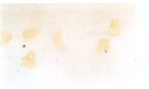

Fig. 1. A photomicrograph showing completely keratinized anuclear epithelial cells present in the cytological sample. No non-keratinized epithelial cells from deeper layers are seen. (x400)
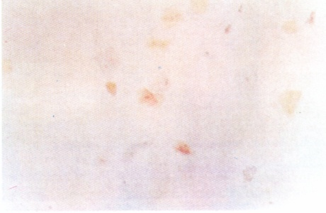
Fig. 2. A photomicrograph showing immature para keratinized cells, with blue cytoplasm and nuclei. The mature para-keratinized cells showed pink / orange cytoplasm. (x160)
Cells in the tape samples occurred either singularly or in groups. When they were in groups, they were frequently arranged in a way that the boundary of one cell was connected that of the adjacent cells and did not overlie it. Therefore it was possible to distinguish the type of cell and to carry out quantification. The cells in all tape samples were stained satisfactorily, the adhesive tape, which formed the background of the sample remained virtually colorless and transparent so that there was no interference from the adhesive tape on the quantification. Inflammatory cells were present in the samples from 4 subjects.
Two subjects were controlled diabetics, one female subject was taking anticoagulants, and three females and three males were smokers. Only one male presented with erythema of the palate. Seven subjects failed to report for the collection of cytological smears at the second stage of the investigation, which was one week after insertion of the new dentures. However, some participants who were unable to attend in one stage of the experiment reported and continued to attend for the remaining stages.
All the subjects had been wearing their old ill-fitting dentures up to the day of insertion of the new dentures.
The degree of keratinization of the tissue was expressed as the percentage of total epithelial cells counted which appeared fully keratinized. The overall level of keratinization ranged from 0 % to 100 %.
Table I present the mean percentage of keratinization found for each male and female at the different sampling periods.
The Mann-Whitney U Test showed a significant difference in keratinization between males and females (Table II), there was also a difference between the keratinization levels at insertion of the new dentures, which showed keratinization from old dentures, and at the various periods of sampling (Table II).
A significant difference (P<0.05) was found in all cases for the females and all but the three-month sampling in the male subjects. Figs 3 and 4 show the change in the mean levels of palatal keratinization in male and female subjects respectively.
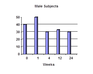
Fig. 3: Changes in mean % keratinization
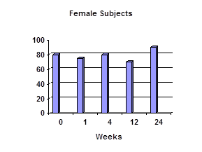 Fig. 4:
 Changes in mean % keratinization
DiscussionThe maintenance of health of the soft tissues beneath a denture base should be of primary importance to the dentist. Although the actual relationship between inflammation within these tissues and resorption of underlying bone has yet to be fully defined, there appears to be an association between the two events. To the present time, clinical observation has been the method by which inflammatory changes within denture bearing mucosa are assessed. More objective methods of assessing epithelial changes could prove to be of use in monitoring mucosal response to dentures. Exfoliative cytology offers a simple, non-invasive technique for examining the maintenance of the stratum corneum, the outermost mechanically protective barrier of the masticatory mucosa.
By assessing the changes in epithelial maintenance, the response of the soft tissues to both the biomechanical features of a denture and to denture plaque control could be monitored. Ideally, the early recognition of epithelial breakdown might allow preventive measures to be undertaken by the clinician to halt progression of the tissue damage and subsequent inflammatory changes.
Unfortunately, there remains a controversy within the literature as to the response of masticatory mucosa to dentures. Some of the studies based on tissues biopsies (2) found increased keratinization of epithelium (2,6) while others found a decrease in keratinization (1,7).
Also some cytological investigations (8) found increased keratinization while others found decreased keratinization (9,10,11). Before such changes can be related to biomechanical features or plaque control the response of mucosa to complete dentures needs to be clarified.
The present study monitored the changes in keratinization of denture bearing tissues in a group of edentulous subjects. The small number of smokers involved in this study did not allow their keratinization level to be analyzed separately. However, both cytological and biopsy studies (9,7) failed to substantiate Markov’s finding of increased keratinization in smokers (8).
The oral cytological smear technique was first proposed by Sandler 1964 (3) and has been used as a technique for the study of variation in oral mucosa keratinization in a number of studies. However, the accepted method of oral cytological sampling has its limitations in that a variation in the depth of the tissue penetrated with the tongue blade may occur depending upon the amount of pressure applied. This would be a disadvantage in studies of the superficial cell keratinization where percentage values of fully keratinized cells are assessed.
In this study we have used Harinasuta’s cytological technique (4), as it offers particular advantage in surveys of the response of supporting mucosa to prostheses because it allows the keratinization level to be studied without interference from epithelial cells from the deeper layers. Even small areas of mucosa can be sampled and provide sufficient numbers of epithelial cells to permit quantification of the level of keratinization. The method may be performed rapidly and offers the possibility of repeated sampling from the same site.
In this study the initial mean levels of keratinization were 41.9 % for males and 81% for females. McMillan found no differences in keratinization levels of males and females in non-denture wearers (11). This was supported by the work of Hairnasuta (4) who found 99 % keratinization in both males and females in a group of young dentate adults. Markov (8) found that keratinization was higher in male subjects but failed to support this statistically. Sandler technique (3) was used to study subjects who had had complete dentures made eighteen months previously and it was found that keratinization was more abundant in men (12).
The results of the present study showed that the level of keratinization decreases in males with the use of dentures, a finding that contradicts previously published data (8). A possible explanation for such a difference may be that the particular denture habits of the subjects may have influenced the results. The role of denture and general oral hygiene would seem to be important (13). Plaque collecting upon the surface of the denture base might irritate the underlying mucosa and initiate a change from ortho keratinization to para keratinization. It could be that the female subjects in our study kept their dentures cleaner than the male subjects. Para functional habits have also been suggested to reflect keratinization (8).
In this study the change in keratinization brought about by the insertion of new complete dentures was examined over a period of six months. It was found that the keratinization levels increased in the female population examined but decreased in the male population. The range of keratinization was from 0-100% throughout the study period. A similar range had been found by other studies that found the most patients to be in the range of 50% keratinization (8). About 58% (10) and 37% (12).
Throughout the six months investigation, periodic increases and decreases in keratinization were seen in individual subjects. While this may reflect a change in their denture wearing habits, levels of keratinization may also have been affected by the general health of the patient.
The factors involved in causing changes of epithelial keratinization in denture supporting mucosa is complex. Some researchers proposed that movement of the denture base during function may be contributory, while others had proposed that denture bases may actually protect tissues especially during the process of mastication (9,14). It was also emphasized that the biomechanical features of the dentures are of paramount importance to the health of the mouth under complete dentures (14).
Conclusion
The wearing of complete acrylic dentures disturbs the denture supporting epithelium of the palatal mucosa and results in decreased levels of epithelial keratinization. The fitting of new complete dentures in a group of subjects brought about further changes in the keratinization levels of palatal tissues, a significant difference was found between the mean levels of keratinization of male and female subjects, and the level of keratinization in individual subjects showed periodic increases and decreases over a six month period. A further study using a greater number of subjects, and in which more attention should be given to defining the denture wearing habits of the subjects and also assessing the biomechanical features of their dentures is indicated.
Table I:The mean percentage keratinization found in male and female subjects.
|
|
Subjects
|
At insertion
|
One week
|
One month
|
Three months
|
Six months
|
|
Male
|
12
|
38.3
|
51.1
|
32.0
|
35.7
|
15.5
|
|
Female
|
13
|
76.0
|
79.2
|
82.5
|
69.2
|
87.0
|
Table II:Comparison between the mean % of keratinization at insertion and at different intervals of samplings
|
Period of samplings
|
Male
|
Female
|
|
No.
|
Mean
|
SD
|
P
|
No.
|
Mean
|
SD
|
P
|
|
0 week
|
12
|
41.9
|
33.7
|
P<0.05
|
13
|
81.1
|
28.0
|
P<0.05
|
|
1 week
|
8
|
51.1
|
44.0
|
P<0.05
|
10
|
78.5
|
25.3
|
P<0.05
|
|
1 month
|
6
|
31.9
|
22.9
|
P<0.05
|
9
|
82.5
|
23.0
|
P<0.05
|
|
3 months
|
5
|
33.7
|
37.4
|
NS
|
10
|
69.2
|
28.1
|
P<0.05
|
|
6 months
|
6
|
30.9
|
36.4
|
P<0.05
|
6
|
87.8
|
11.9
|
P<0.05
|
References
1. Ostlund SGS. The effect of complete dentures on the gum tissues. Acta Odont Scand 1958; 16: 1-36.
2. Kapur K. The effect of complete dentures on alveolar mucosa. J Prosth Dent 1963; 13: 1030-1037.
3. Sandler HC. Reliability of oral exfoliative cytology for detection of oral cancer. J Amer Dent Ass 1964; 68: 489-493
4. Harinasuta S. Development and assessment of a sampling technique for epithelium for prostheses. M. Sc. Thesis, University of London, 1986.
5. Papanicolaou GN. A new procedure for staining vaginal smears. Science 1942; 95: 438-439
6. Jani RM, Bhargava K. A histological comparison of palatal mucosa before and after wearing complete dentures. J Prosth Dent 1976; 36: 254- 260.
7. Watson IB, MacDonald RD. Oral mucosa and complete dentures. J Prosth Dent 1982; 47: 133-135.
8. Markov NJ. Cytologic study of keratinization under complete dentures. J Prosth Dent 1968; 20: 8-13.
9. Mi Ani S, Shakler G, Yurkstatas AA. The effect of dentures on the exfoliative cytology of palatal and buccal oral mucosa. J Prosth Dent 1966; 16: 513-521.
10. Lindholm K, Hakala PE, Makila E. Leukocyte count, and keratinization of the palatal denture bearing mucosa. J Prosth Dent 1982; 47: 440 – 444.
11. Akal UK, Aydogan S, Oygur T, et al. Keratinization of palatal mucosa beneath metal – based removable partial and acrylic-based complete dentures compared with normal palatal mucosa; a clinical, cytological and histological study. J Marmara Univ Dent Fac 1997; 2(4): 665-672.
12. McMillan DR. The cytological response of palatal mucosa to dentures. Dent Pract 1972; 22: 302 – 304.
13. Theilade E, Budtz-Jorgensen E, Theilade J. Microflora of denture plaque in denture induced stomatitis. J Dent Res (Supplement) 1985; 64: 682-685. (Abstract No. 180).
14. Markov NJ. Cytologic study of the effect of some biomechanical principles of complete denture construction on keratinization of the mucosa of the edentulous ridge. J Prost Dent 1969; 21: 132-137.