Objective: To report the clinical efficacy of external fixator used for the management of gunshot induced open fractures and to report our experience at King Hussein Medical Center.
Methods: The medical records of 60 patients with open gunshot extremity fractures, treated at King Hussein Medical Center from January 1999 to January 2004, were reviewed retrospectively. Only patients with diaphyseal comminuted fractures were included in this study. All patients had debridement, antibiotic therapy, and stabilization of the fracture with an external fixator (either the self-made external fixator using plastic tube and bone cement or the AO tubular system). The external fixator was left in place for 6-54 (average 16) weeks. Clinical follow up ranged from 18-36 months after operation with an average of 26 months.
Results: Low-velocity fractures occurred in 25 patients. High-velocity fractures occurred in 35 patients. All fractures were open, comminuted and displaced. Eight cases of nerve injury were seen (13%). Arterial injuries were present in 9 patients (15%). Clinical evidence of wound infection was documented in the records of 14 patients (23%) including pin tract infections (10%). The tibia and femur were the most frequent sites accounting for 11 infections, with only three located in the upper extremity. Three patients had septicemia and four had chronic osteomyelitis. The wounds healed by granulation tissue in 47 patients; seven had secondary closure, and four had split skin grafts, two had free flaps.
Conclusion: The complications associated with gunshot injuries could be reduced to the minimum if the basic principles of management are strictly adhered to. External fixation is the preferred initial treatment for stabilizing severe open missile fractures. We recommend this treatment for low-velocity and high- velocity gunshot open fractures.
Key words: Gunshot open fractures, External fixator, Treatment.
JRMS April 2007; 14(1): 17-21
IntroductionComplex open fractures from gunshot wounds associated with neurovascular injuries present a therapeutic challenge to the orthopaedic surgeon. The fractures associated with these injuries are often comminuted and unstable. Bone loss is common. Soft tissue disruption plays a more important role in high-energy gunshot-induced fractures. In these cases, external fixation is the treatment of choice for stabilization.
Gunshot injuries have become increasingly common in most countries particularly the United States of America(1). Many papers found in the literature about gunshot injuries are from the USA(2-4).
The complications associated with these injuries tend to be related to the degree of energy transfer(5) and include wound infection, neurovascular injury, compartment syndrome, delayed union and non-union of fractures.
The extent of tissue damage inflicted by a bullet is influenced by the bullet's velocity, weight, size, shape, and tumbling characteristics as well as by the character of the tissue that it strikes(6). The capacity for wounding increases exponentially as velocity increases (KE = 1/2 mv², where KE is kinetic energy, m is the mass and v² represents the speed).
Bullets from most low-velocity weapons used in a civilian setting travel at an average of 1000 to 2000 feet (305 to 610 meters) per second, while bullets from high-velocity weapons exceed 2000 feet (610 meters) per second.
Most military ammunition travels at 2400 to 2900 feet (732 to 884 meters) per second and that from M-16 rifles travel at 3250 feet (991 meters) per second(6). It is only when the impact velocity is greater than 600 feet (183 meters) per second that shock waves from the missile have much effect.
Temporary cavitation becomes apparent when velocity exceeds 1000 feet (305 meters) per second and becomes extensive when velocity exceeds 2000 feet (610 meters) per second(7). Damage to long bones depends, to some extent, on the area of involvement. When a low-velocity bullet strikes the cancellous portion of the metaphysis of a bone, a so-called drill-hole defect frequently results with little or no comminution. Conversely, when the same bullet strikes the shaft, it often produces a comminuted fracture with so-called butterfly fragments at right angles to the wound track(8,9) (Fig. 1).
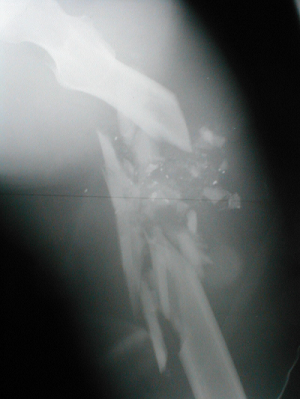 Fig. 1:
 High velocity, severely comminuted gunshot fracture of the Femur.
There is still much debate over the treatment of fractures caused by gunshot. A wide range of methods ranging from the non-operative such as low-tech splintage through external fixation to internal fixation or intramedullary nailing have been advised(10).
The purpose of this study was to report on our experience in treating gunshot open fractures with external fixators (Fig. 2).
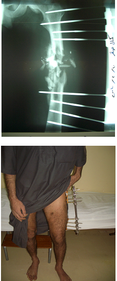
Fig. 2: High velocity femoral fracture initially treated by an AO tubular External Fixation. Same patient with the external fixator which has been applied to him.
Methods
The medical records of 60 patients with open gunshot extremity fractures, treated at King Hussein Medical Center from January 1999 to January 2004, were reviewed retrospectively. A total of 60 patients with long bone fractures were identified. The mean age of patients was 24 years (range 18-38). All patients were males. Thirty-three patients were military and 27 patients were civilians.
The etiology of trauma was accidental in 18 cases, an assault in 31, self-inflicted in six and unknown in five instances. Patients were admitted to the hospitals within few hours of the injury. The wounding agent was a pistol in half of the cases and a rifle and shotgun in the remaining half.
The etiology and trauma mechanism were studied. Data collected also included age, gender distribution of the victims, location of the wounds, fracture type, early/late treatment of the patients, and postoperative complications.
The injuries were divided into high- and low-velocity based on a combination of history, clinical assessment of the wound and radiographic findings.
The majority of cases were seen within six hours of the injury and tetanus toxoid was administered if the patient was unsure history of immunization, or had not received a booster over the last ten years.
Low-velocity fractures occurred in 25 patients. The Low-velocity wounds were generally managed by minimal wound excision and cleansing.
High-velocity fractures occurred in 35 patients. All High-velocity wounds were surgically explored; skin edges and wound track excised with debridement of necrotic soft tissues, bone fragments were irrigated by copious amount of normal saline and kept attached to soft tissues, deperiostised burned fragments and those not attached to soft tissues were excised. All patients had antibiotic therapy and stabilization of the fracture with an external fixator (either the self made external fixator using plastic tube and bone cement or the AO tubular system). Only patients with diaphyseal comminuted fractures were included in the study. Surgical exploration was undertaken for wounds with concomitant neurovascular injuries.
Clinical follow up ranged from 18-36 months after operation with an average of 26 months (Fig. 3).
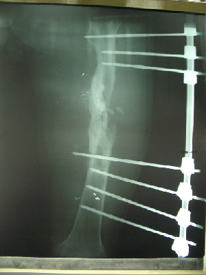 Fig. 3:
 External Fixation with bone consolidation. In this diaphyseal comminuted femoral fracture. The fractures healed after wound debridement.
Results
Low-velocity fractures occurred in 25 patients. High-velocity fractures occurred in 35 patients. Clinical evidence of wound infection was documented in the records of 14 patients (23%) including pin tract infections (10%). All wound infections were predominantly in high-velocity injuries. The tibia and femur were the most frequent sites accounting for 11 infections, with only three located in the upper extremity. Three patients had septicemia and four chronic osteomyelitis.
All fractures in the study were open comminuted and displaced. The location of the fracture was the humerus in 12 (20%) cases, radius in nine cases (15%), femur in 11 cases (18.3%) and tibia in 28 cases (46.7%) (Table I). Eight cases of nerve injury were seen (13.4%). One sciatic (1.7 %), three radial (5%), two peroneal (3.3%), one median (1.7%) and one (1.7%) ulnar were identified (Table II). Five recovered spontaneously while others required grafting.
Arterial injuries were present in nine patients (15.1%). The injury was caused by rifles (4), shotguns (2), and pistols (3). The injured arteries were brachial three cases (5%), radial one case (1.7%), femoral two cases (3.3%), popliteal one case (1.7%), anterior tibial one case (1.7%) and posterior tibial one case (1.7%1) (Table III). The arteries were repaired by end-to-end anastomosis or vein patch. Fasciotomy was done routinely after vascular repair or when compartment syndrome was suspected (three cases).
The wounds healed by granulation tissue in 47 patients; seven had secondary closure, four had split skin grafts and two had free flaps. The external fixator was left in place for 6-54 weeks (average 16 weeks) (Fig. 4).
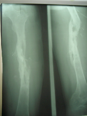 Fig. 4:
 Femur after removal of external fixation, with good bone consolidation, and complete bone healing.
Bone consolidation was completed within 6-54 weeks (average 16 weeks), in 49 patients (81.8%). Seven patients had delayed union (11.6%) for which external fixators were left in place for 26 weeks in those patients until complete bone consolidation occurred.
Four patients had non-union (6.6%). Non-union cases were treated by internal fixation and bone grafting, all of them eventually healed.
Table I: Frequency of bone involvement in gunshot injuries
|
Bone involved
|
No.
|
%
|
|
Humerus
|
12
|
20
|
|
Radius
|
9
|
15
|
|
Femur
|
11
|
18.3
|
|
Tibia
|
28
|
46.7
|
|
Total
|
60
|
100
|
Table II: Frequency of nerve injury in gunshot injuries.
|
Nerve injured
|
No.
|
%
|
|
Sciatic
|
1
|
1.7
|
|
Radial
|
3
|
5
|
|
Peroneal
|
2
|
3.3
|
|
Median
|
1
|
1.7
|
|
Ulnar
|
1
|
1.7
|
|
Total
|
8
|
13.4
|
Table III: Incidence of artery injury in gunshot injuries
|
Artery Injured
|
No.
|
%
|
|
Brachial
|
3
|
5
|
|
Radial
|
1
|
1.7
|
|
Femoral
|
2
|
3.3
|
|
Popliteal
|
1
|
1.7
|
|
Anterior
tibial
|
1
|
1.7
|
|
Posterior
tibial
|
1
|
1.7
|
|
Total
|
9
|
15.1
|
Discussion
This study of treatment for gunshot fractures was conducted to identify the specific problems of firearm injuries and their treatment with external fixator.
The destructive force of gunshots is directly proportional to the amount of energy released into surrounding tissues. Wang et al(11) described three zones of injury secondary to missiles: (1) primary wound tract/permanent cavity; (2) a contusion zone of muscle adjacent to missile tract; and (3) a concussion zone (outside congestion in varying degrees).
In civilian gunshot wounds, the damage is essentially confined to the bullet tract because of the relatively low-velocity of handgun ammunition(3,6,12). Military weapon velocities are in excess of 3000 ft/second. The explosive characteristics result from the expansion of a temporary cavity exceeding the elastic capacity of the surrounding tissues. Bullets are not sterilized by firing(13), and fractures associated with gunshot wounds are open, communicating with the external environment. Therefore, all gunshot fractures are contaminated and potentially infected(6).
Shotgun was the causative agent in about 12% of fractures. Range is the most important determinant of the amount of damage inflicted by a given shotgun charge. The second one is the type of pellet(14). In our study, most of these wounds occurred at a range closer than 5 meters. Most pellets were about 2.5 mm in diameter. Therefore, most of these injuries were severe with massive soft tissue devitalization.
When pellets, wadding, gunpowder debris, and casing debris are blasted into an extremity with other foreign bodies and massive bacterial contamination, these wounds may even be “worse” than large so-called high-velocity wounds)15(. Our rate of infection was generally high. An infection rate of less than 4% has been reported for low-velocity injuries(16). Our rate of 16% for low-velocity injuries despite routine antibiotic administration indicates a need for greater attention to wound care. Ten out of thirty-five of our high velocity injuries were complicated by infections. In retrospect and in keepind with the general principles of management of high-velocity injuries(2,17,18), most of these wounds should have been more thoroughly debrided and closely followed-up to decrease the incidence of infection.
With respect to associated neurovascular injuries in our series, the rate was comparable to those in the literature(2,15). This is due to relatively greater number of high-velocity injuries in our patients.
Many authors have reported excellent results in treating complicated fractures by external fixator, and many agree that it is the method of choice in the management of missile injuries(19-22). This technique has many advantages, especially in the immobilization of gunshot fractures, including large soft-tissue lesions. All of our cases were managed by external fixation with good results.
Conclusion
The complications associated with gunshot injuries can be reduced to the minimum if the basic principles of management are strictly adhered to. The external fixation is the preferred form of primary stabilization of severe open fractures. The treatment of the soft tissues, skin coverage, and ultimately reconstructive surgery are facilitated. Early limb function allows for a better final result. For delayed union and pin-tract infection a different definitive method, such as internal fixation or a plaster case, is required. External fixation is the preferred initial treatment for stabilizing severe open missile fractures. We recommend this treatment for low-velocity and high- velocity gunshot open fractures.
References
1.
Hull JB. Management of gunshot fractures of the extremities. J Trauma 1996; 40(3): 193-197.
2.
Billings JB, Zimmerman MC, Aurori B, et al. Gunshot wounds to the extremities: Experience of a level I trauma center. Orthop 1991; 20: 519-524.
3.
Marcus NA, BlairWF, Shuck JM, Omer GE. Low-velocity gunshot wounds to extremities. J Trauma 1980; 20: 1061-1064.
4.
Ordog GJ, Wasserberger J, Balasubramaniam S, Shoemaker W. Civilian gunshot wounds-outpatient management. J Trauma 1994; 36(1): 106-111.
5.
Bowyer GW, Rossiter ND. Management of gunshot wounds of the limbs. J Bone Joint Surg (Br) 1997; 79(6): 1031-1036.
6.
Howland WS, Ritchey SJ. Gunshot fractures in civilian practice. An evaluation of the results of limited surgical treatment. J Bone and Joint Surg 1971; 53-A: 47-55.
7.
Hennessy J, Banks HH, Leach RB, Quigley TB. Extremity gunshot wound and gunshot fracture in civilian practice. Clin Orthop 1976; 114: 296-303.
8.
DeMuth WE. Bullet velocity and design as determinants of wounding capability: An experimental study. J Trauma 1966; 6: 222-232.
9.
DeMuth WE, Smith JM. High-velocity bullet wounds of muscle and bone: The basis of rational early treatment. J Trauma 1966; 6: 744- 755,
10.
Apley AG, Solomon L. Principles of fractures. In: Apley AG, Solomon L, editors. Apley's system of orthopedics and fractures. 6th edition. London: Butterworth Scientific 1982; 333-368.
11.
Wang Z, Feng JX, Liu YQ. Pathomorphological observation of gunshot wounds. Acta Chir Scand 1982; 508:185-186.
12.
Dugas R, D’Ambrosia R. Civilian gunshot wounds. Orthopedics 1985; 8(9): 1121-1151.
13.
Thoresby FP, Darlow HM. The mechanisms of primary infection of bullet wounds. Br J Surg 1967; 54: 359-360.
14.
Wilson JM. Shotgun ballistics and shotgun injuries. West J Med 1978; 129: 149-150.
15.
Tikka S, Bostman O, Martinen E, Makitie I. A retrospective analysis of 36 civilian gunshot fractures. J Trauma 1996; 40(3): 212-216.
16.
Woloszyn JT, Uitvlugt GM, Castle ME. Management of civilian gunshot fractures of the extremities. Clin Orthop 1988; 226: 247-251.
17.
Coupland RM. Technical aspects of war wound excision. Br J Surg 1989; 76: 663–667.
18.
Qidwai S, Khattak Z, Malik M. Management of gunshot injuries to the limbs. SMJ 1999; 20(8): 587-593.
19.
Coupland RM. War wounds of bones and external fixation. Injury 1994; 25:211–217.
20.
Dubravko H, Zarko R, Tomislav T, et al. External fixation in war trauma management of the extremities-experience from the war in Croatia. J Trauma 1994; 37(5): 831-834.
21.
Davila S, Mikulic D, Davila NJ, et al. Treatment of war injuries of the shoulder with external fixators. Mil Med 2005; 170(5): 414-417.
22.
Miric D, Bumbasirevic M, Radulovic N, Lesic A. External fixation of war injuries of the proximal femur. Acta Chir 2005; 52(2):101-105.