Objective: To define the clinical spectrum of tracheobronchial foreign body aspiration in adults, evaluate the efficacy of bronchoscopy, and determine outcome and complications at King Hussein Medical Center.
Methods: Retrospective analysis of 33 consecutive clinical series from a referral-based medical center (King Hussein Medical Center) was conducted. Case records of 33 adult patients (over 14 years of age) suspected to have foreign body aspiration over the past eight years were analyzed. All had bronchoscopic evaluation. Clinical-radiological features, types and location of foreign body were studied. About 98% of patients had their foreign bodies identified and removed using Flexible Fiberoptic Bronchoscopy while rigid bronchoscopy was needed in only three (9%) cases.
Results: Of 33 consecutive patients, 25 patients (76%) were females and the majority was below 30 years of age. A total of 30 patients (91%) presented within the first 24 hours after foreign body inhalation. A radio-opaque foreign body was evident in chest X-rays of 85% of the patients. Pins used to fix head scarves constituted 64% of the foreign bodies removed. The rest included a piece of bone, food particles and seeds. A total of 29 patients (88%) were discharged on the same day of the procedure.
Conclusions: Ttracheobronchial foreign body aspiration in adults can occur in various settings. High clinical suspicion is necessary for diagnosis. Removal of foreign bodies can usually be accomplished with fiberoptic bronchoscopy. In our series the most commonly removed foreign bodies were metallic pins.
Key words: Foreign body aspiration, Fiberoptic Bronchoscopy
JRMS Dec 2007; 14(3): 12-14Introduction Foreign body (FB) aspiration is an important cause of adult morbidity and a rare cause of mortality, particularly in early adulthood and elderly. It is potentially life-threatening event and may also cause chronic lung injury, if not properly managed.(1,2) The symptoms and signs can be confused with those of asthma, and the X-Ray findings with those of pneumonia. Foreign bodies may cause chronic pulmonary infections, bronchiectasis and lung abscess.(2-4) An early diagnosis and management of the patient with an inhaled FB offers a diagnostic challenge to the physician. Flexible Fiberoptic Bronchoscopy (FFB) proved to be a useful tool in diagnosis and management of such patients. This study reviews the clinical data of adult patients presented to King Hussein Medical Center (KHMC) with suspected FB inhalation.
Methods The medical records of all adults presented with suspected foreign body aspiration to KHMC, between June 1998 and June 2005, were analyzed retrospectively. The following data were collected: sex, age, residence, duration of illness, availability of definitive history, bronchoscopic procedure, associated findings on bronchoscopy, number, type and location of the foreign body, duration of hospital stay after its removal, and complications. All patients suspected of foreign body aspiration were subjected to fiberoptic bronchoscopy under local anesthesia and mild sedation using intravenous midazolam in a dose of 0.05-0.20mg/kg body weight and local xylocaine 2% spray in the tracheobroncheal tree. Patients in whom no foreign body was found were excluded from final analysis.
Results Thirty-seven adults were presented with suspected foreign body aspiration during this period. No foreign body was found in four subjects and they were excluded from the analysis. Of the 33 patients with foreign body aspiration, eight (24%) were males and 25 (76%) were females. Most patients (n=27, 82%) were below 30 years of age; the median age (range) was 16 years (14–42). Thirty patients (91%) reported within 24 hours of the event. The median duration of symptoms prior to presentation was 12 hours. There was a delay of more than two weeks in three (9%) cases. A definite history of choking following foreign body aspiration was present in 31 (94%) cases. Chief clinical features are shown in Table I.
Table I: Clinical features of patients with foreign body aspiration
|
Feature
|
No
|
%
|
|
Choking
|
31
|
94
|
|
Paroxysmal
cough
|
32
|
97
|
|
Decreased
air entry
|
14
|
42
|
|
Wheezing
|
11
|
33
|
|
Fever
|
4
|
12
|
Chest radiographs were helpful in providing evidence of possible foreign body aspiration in 30 (91%) patients. A radio-opaque foreign body was evident in 28 cases (85%). Radiological features of air trapping were seen in 20 (61%) cases, while collapse/atelectasis was seen in four (12%) cases. Mediastinal shift was seen in 6% cases only. Normal radiograph or one showing non-specific findings was present in 8% of cases. Details of treatment received prior to referral were available in only seven cases. The treatment received included antibiotics, intravenous fluids, oxygen supplementation and bronchodilators.
Bronchoscopy showed tracheobronchial edema or granulation tissue (40%), inflammation (30%) and purulent secretions in 15% of cases. A single foreign body was present in 32 (97%) cases. In 73% of cases, a FB was impacted in the intermediate bronchus. The location of foreign bodies is shown in Table II. Foreign bodies removed were pins in 21 (64%) of cases, pulses and seeds in four cases, a piece of plant leaf in two cases, a piece of bone in two cases, and food particles in four patients. Some of these FBs are shown in Fig. 1.
Table II: Foreign body location
|
Location
of Foreign body
|
No
|
%
|
|
Trachea
|
1
|
3
|
|
Carina
|
2
|
6
|
|
Intermediate
bronchus
|
24
|
73
|
|
Right
Main bronchus
|
4
|
12
|
|
Left
main bronchus
|
2
|
6
|
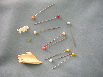 Fig 1:
 Some of the foreign bodies removed by bronchoscopy
A total of 29 patients (88%) were discharged within the same day of FB removal while five patients stayed in hospital for more than 48 hours because of complications like fever and bronchospasm. Two patients with persistent symptoms required a repeat bronchoscopy and showed edema and secretions with no remaining foreign bodies. No deaths were reported in all cases dealt with after bronchoscopy.
Discussion Tracheobronchial FB aspiration is an important life threatening condition in all age groups and particularly in younger children. The anatomic relation of the larynx, shouting, crying and playing while eating contribute to this hazard. Most patients in our study were below 30 years of age, which is similar to that reported in other series.(5,6) The female-male ratio in our study was 2:1, which differs from some similar other studies.(5-7) This might reflect local cultural habits among the studied population of our study. The majority of FBs removed were pins used by young females to fix their head scarf, which were inhaled when they put a pin in their mouth while fixing their head scarves. The fact that about 9% of patients arrived at the hospital more than two weeks after inhalation is of some concern, bearing in mind that a positive history of aspiration was obtained in 94% of patients. Some were treated with antibiotics and bronchodilators, before referral with suspected foreign body. One patient was referred more than seven days after onset of symptoms. Medical staff had overlooked the significance of choking when the initial assessment was made. Factors, which may delay diagnosis include: wrong/incorrect diagnosis, abscence of symptoms (particularly after the acute initial phase of dyspnea), and diverse clinical features. Common features included choking, paroxysmal cough and decreased air entry.
Chest radiograph is important for diagnosis. Since the most commonly aspirated objects are radioopaque pins, their presence is usually established by chest X-ray in 85% of patients. Indirect signs of atelectasis or air trapping due to partial obstruction were demonstrated in 15% of the patients. In other studies a normal chest radiograph was reported in 9-30% of cases.(4,8,9)
Radiographs were normal in 15% of our cases. The presence of x-ray abnormality is related to size, type, shape and location of FB, and the pattern and extent of bronchial obstruction. In more than half of the cases, the FB was lodged in the right main bronchus. This is in agreement with the findings of other studies,(1,9,10) and is probably determined by anatomical factors.
There were no deaths in this study reflecting the safety of bronchoscopic removal around the world. It is recommended that patients satisfying the following criteria should be subjected to bronchoscopy: history of definite or suspected FB aspiration; features of foreign body aspiration, e.g. choking, wheezing, stridor and paroxysmal cough; recurrent chest infections with no apparent cause, when bronchial asthma has been excluded; and chest radiograph suggestive of foreign body aspiration. FFB is a safe and effective way of foreign body removal from the tracheobronchial tree.
References1.
Toker A, Tanju S, Dilege S, Kalayci G. Foreign body aspiration: Where can it be? Eur J Cardiothorac Surg 2003; 24(2): 301-302.
2.
Dilege S, Toker A, Tanju S, Kalayci G. An unusual intrapleural foreign body: Ignored aspiration. Eur J Cardiothorac Surg 2002; 21(3): 593-594.
3.
Yang TL, Hsu MM. Occult foreign body aspiration in adults. Otolaryngol Head Neck Surg 2003; 128(1): 154-156.
4.
Yilmaz A, Akkaya E, Damadoglu E, Gungor S. Occult bronchial foreign body aspiration in adults: Analysis of four cases. Respirology 2004; 9(4): 561-563.
5.
Baharloo F, Veyckemans F, Francis C, et al. Tracheobronchial foreign bodies: Presentation and management in children and adults. Chest 1999; 115(5): 1357-1362.
6.
Swanson KL, Edell ES. Tracheobronchial foreign bodies. Chest Surg Clin N Am 2001; 11(4): 861-872.
7.
Debeljak A, Sorli J, Music E, Kecelj P. Bronchoscopic removal of foreign bodies in adults: Experience with 62 patients from 1974-1998. Eur Respir J 1999; 14(4): 792-795.
8.
Dikensoy O, Usalan C, Filiz A. Foreign body aspiration: clinical utility of flexible bronchoscopy. Postgrad Med J 2002; 78(921): 399-403.
9.
Demircan S, Celik B, Basoglu A. An unusual history of a foreign body aspiration. Asian Cardiovasc Thorac Ann 2004; 12(4): 387-388.
10.
Herget E, Hiorns MP, Mayo JR. Foreign-body aspiration as a cause of an asymmetric carina: Case report. Can Assoc Radiol J 2004; 55(1): 13-15.