ABSTRACT
Objectives: To investigate our experience with “Neck Advancement” as a useful technique in late reconstruction of some neck and lower face post-burn scars in addition to evaluate the limitations and complications of this technique.
Methods: During the period between 1999-2006, a total of 110 “Neck Advancement” procedures were performed on 57 patients with mild to moderate neck and lower face scars at the Royal Jordanian Rehabilitation Center. Forty one of these 57 patients had previous scar revisions or serial excisions ranging from 1-6 procedures per patient prior to the neck advancement. The medical records, pre- and postoperative photographs of these patients were retrospectively studied in terms of age, gender, cause of burn, duration since the acute burn, previous attempts at reconstruction, indications for surgery, results, and postoperative complications.
Results: The range of neck advancements was 1-4 procedures per patient. The overall post operative improvement was good in 45 (79%) patients, satisfactory in 9 (16%) patients, and poor in only 3 (5%) patients. There were a total of 13 complications among our group of patients. Apart from simple wound problems related to this technique, the main complication was facial or neck asymmetry, a pseudo-torticollis that tends to occur with unilateral advancements.
Conclusion: We found this technique useful in the late reconstruction of a large number of patients with mild to moderate facial and neck burns. It is easy, quick, repeatable and convenient to the patient with a low frequency of complications. It is an additional technique to the armamentarium of the reconstructive surgeon to deal with such difficult burns. It has limitations and is not suitable for the more extensive neck burns.
Key words: Advancement, Burn, Face, Late Reconstruction, Neck
JRMS April 2009; 16(1): 43-50IntroductionFacial and neck burns represent between one fourth and one third of all burns.(1) The most vulnerable groups to burn injury are the very young, the elderly, and the physically handicapped.
The treatment of burn defects is a challenging enterprise for the reconstructive surgeon. Burns of the head and neck present a particularly complex problem, as the defects involve critical and highly visible structures that are important functionally and cosmetically. In addition, burns of the face and neck tend to be extensive and often involve adjacent tissues, limiting reconstructive options.
The neck is susceptible to unfavorable post-burn scar outcomes. Second and third-degree burns that require healing by secondary intention or excision and grafting are associated with poor scar outcomes(2-5) particularly with simple serial excisions.
Burn reconstruction may be characterized as a battle between favorable and unfavorable scars. More frequently than not, aesthetic burn scar revision entails converting a "patch" scar into a linear one. Large areas frequently need to be resurfaced, preferably putting the seams at the junction of aesthetic units if possible.(6) Patients with old burn scars of the face and neck are among the most difficult to treat. All current techniques are to a large extent unsatisfactory to the patient with regard to aesthetic improvement. Although different methods of reconstruction are used for the treatment of neck scars, including the use of split-thickness skin graft, local flaps, expanded local flaps, thin pedicled flaps, free flaps, and expanded free flaps, a limited and unsatisfactory outcome often results.(7-13)
This study was conducted in order to review the experience gained and the difficulties encountered in the reconstruction of the post-burn face and neck scars utilizing, for want of a better word, “neck advancement techniques”. This utilizes some of the well established neck and facelift techniques used by the aesthetic plastic surgeon in rejuvenating the aging neck and face and applying these same principles with appropriate modifications to the more challenging scenario of reconstructing the burnt neck and face which are often disfigured and often contracted and where other techniques were deemed either inappropriate or unjustified. The technique we used was adapted from some of the teachings of Feldman(13) on the subject.
The main objective of the study was to evaluate the outcome of such techniques and assess the complication rates and repeatability of the operation on the same patient and whether the extensive undermining involved achieves better results because of the decreased tension on the suture line, than simple serial excisions with limited undermining or tissue expansion as was usually performed for such cases. The high recurrence rate of serial excisions and the high complication rates of tissue expansion in the neck are the main reason for
us to seek other alternatives for the reconstruction of these burn scars.
MethodsFrom June 1999 to October 2006, 57 patients with mild to moderate neck and lower face post burn scars were treated at the Royal Rehabilitation Center utilizing neck advancement techniques. The medical records, pre- and postoperative photographs of these patients were retrospectively studied in terms of age, sex, cause of burn scar, duration since the acute burn, previous attempts at reconstruction, indications for surgery, results, and postoperative complications. Follow up ranged from 6 months to 7.5 years.
There were twenty six male patients and thirty one female patients in the group (M: F=1:1.2). The mean age of the patients was 23.7 years (range 9 to 44 years). Causes of the burn scars were scald burns in twenty nine patients, flame burns in twenty three patients and chemical burn in four patients and an electrical burn in one patient. The percentage of total body surface area burnt was classified into minor (<10%), moderate (10-30%) and severe (>30%).
The demographic data of the cohort of patients is summarized in Table I.
Forty-one of these 57 patients had unsatisfactory previous surgeries for scar revisions and serial excisions ranging from one to six surgeries per patient with a total of 125 procedures (Table II). Hence the reason for adopting a neck advancement technique as an alternative to simple scar revision or stage excision.
The results of the surgery were subjectively evaluated by the surgeon and classified as poor, satisfactory and good in terms of the expectation of the surgery. No objective criteria for assessment were utilized since this was a simple descriptive study and the cohort of patients was very variable in terms of severity of scars, depth of scarring, degree of functional deficit, surface area involved and exact anatomical site.
Indications for Neck Advancement Techniques
We used this technique for mild to moderate burn scars of the neck and lower face where a staged serial excision had either failed or deemed to fail because of the relative width of the scar (Fig.1a).
Table I:Demographic and clinical characteristic data of the study group
|
|
|
n =57
|
|
Age(yrs)
|
Mean
|
23.7
|
|
(Range)
|
9-41
|
|
Gender
|
M:F
|
26:31(1:1.2)
|
|
Cause of burn
|
Scald: Direct Flame: Others
|
29:23:5
|
|
TBSA%
|
Minor: Moderate: Severe
|
37:18:2
|
|
Original Burn Management of Neck
|
Spontaneous Healing: Grafted
|
42:15
|
Table II: No. of scar revisions per patient prior to neck advancement
|
No of
Scar Revisions/ Pt
|
No. of
Patients (%)
|
No of
Surgeries (%)
|
|
0
|
16 (28)
|
0 (0)
|
|
1
|
7 (12.5)
|
7 (5.5)
|
|
2
|
11 (19)
|
22 (17.5)
|
|
3
|
8 (14)
|
24 (19.5)
|
|
4
|
6 (10.5)
|
24 (19.5)
|
|
5
|
6 (10.5)
|
30 (24)
|
|
6
|
3 (5.5)
|
18 (14)
|
|
Total
|
57 (100)
|
125 (100)
|
The technique was also applied where there was hesitancy in the use of tissue expansion in the neck because of the high rate of expander exposure complicating the use of expanders in the neck as noticed from our own experience.
Pre-requisites for the success of the procedure are the presence of a good amount of healthy pliable lower neck and anterior chest wall skin and at most a mild neck contracture. This technique is not suitable for patients with either extensive neck burns or severe contractures. In these situations other established techniques are usually performed such as skin grafts, local pedicled flaps or free flaps.(7- 13)
A good knowledge of facial and neck anatomy, an understanding of geometry and skin biomechanics, a good sense of aesthetics and experience with the limitations of this procedure will avoid some of the complications and prevent the overuse of this technique in inappropriate situations where a more difficult reconstructive option is warranted.
Surgical technique:
The surgical technique we employ has gradually evolved over the last seven years as experience was gained with this procedure. All patients were operated upon under general endotracheal anesthesia, the tube being placed either orally or nasally, depending on the need for the mouth to be free of impediments for surgical requirements. The position of the patient at the commencement of surgery was supine with the neck hyperextended to exaggerate the contracture if any. The planned surgical field is infiltrated with an adrenaline solution in a dilution of 1:100000 to minimize blood loss.
Many of these patients require more than one stage of advancement to achieve a satisfactory outcome. The incision was therefore usually made just above the lower margin of the burn scar and within the scar (Fig 1b); to minimize the use of normal healthy skin for the later suturing of the advancing skin front so as to safeguard valuable healthy skin for the final stage of reconstruction as a precaution for the development of hypertrophy at the suture line.
Once a healthy plane is reached the dissection of the unburned remaining neck skin would proceed in a supra platysmal plane (Fig.1c) all the way down to the infra clavicular region and onto the upper chest wall for about 3-5cm to release all the attachments of the lower neck skin to the anterior chest wall, to recruit more healthy skin from the area and to maximize the release of any tension on the suture line (Fig.1d). Skin undermining is never performed lateral to the posterior borders of both sternocleidomastoid muscles to preserve the nerve and blood supply to the cervical flap. The skin flaps were then mobilized in an upward, lateral or medial direction according to the location of the burn scar on the neck and/or face (Fig.1e).
Burn Scar excision was only performed after advancement. The maximal area of scar that could be excised was estimated by re-draping the mobilized healthy skin over the scar using temporary stay sutures and marking the area ofplanned excision with a marking pen taking into consideration the need
to either maintain or reconstitute a cosmetically acceptable
cervico-mental angle at the level of the hyoid bone.
The marked scar was
then excised starting from the area of minimal tension towards the area
of increasing tension. Before completing the excision and depending on
the amount of release achieved through excision of scar in areas of
increasing tension, draping and marking were repeated, using temporary
stay sutures, to correct for any over or under-estimation in our
original plan of scar excision. Any underlying scar tissue in the
subcutaneous plane was excised down to a healthy plane to release any
contracture bands (Fig.1f).
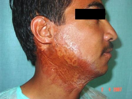
Fig. 1a. Neck & Lower face scar (Preop).
Incision
↓
SCM→ ←Adv.
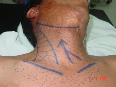
Fig.1b. Planned Incision.
Dots represent extent of undermining.
Fig.1c. Plane of Flap elevation.
Fig.1d. Completed flap elevation.
Fig.1e. Flap advancement.
Fig.1f. Scar excision.
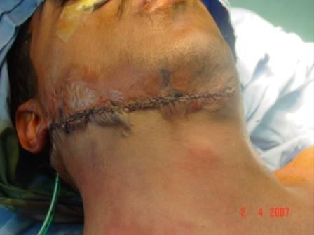
Fig.1g. Final closure over drains
The upper end of the wound is never undermined to avoid any pull or distortion of facial landmarks. The cervico-mental angle is reconstituted if disturbed by tacking sutures of the advanced neck skin to the underlying hyoid bone.
The stay sutures were then removed and hemostasis established. Negative suction drains were used to drain the flap. The advanced neck flap was then sutured in the new position with an air tight two layer wound closure, using fine absorbable subdermal sutures and non-absorbable stitches for the skin (Fig.1g).
The wound is covered with steristrips and the neck is left exposed at all times post-operatively for the early detection of any collection or skin necrosis. One important aspect, which we learned with experience, is to carry the undermining to both sides of the neck even in unilateral burns, to avoid asymmetrical pull on one side which occurred in some of our earlier cases, a complication similar to torticollis. (Fig 2a, b).
The technique with modifications in incision, undermining and direction of advancement can be applied to central neck scars (Fig 3a,b), lateral neck scars, cheek scars (Fig 4a,b) and chin and perioral scars (Fig 5a,b). We have also used this technique in other patients, not included in this study, with giant nevi of the face and neck, traumatic scars and residual scars of vascular lesions.
ResultsA total of 110 neck advancement procedures were performed on 57 patients with mild to moderate neck and lower face post-burn scars. The range was 1-4 procedures per patient (Table III).
Indications for surgery were for aesthetic purposes in 42 patients (74%), the presence of discomfort associated with the scar in 11 patients (19%), and limitation in active or passive range of motion of the neck in four patients.
The range of neck advancements was 1-4 procedures per patient. The overall post operative improvement was good in 45 (79%) patients, satisfactory in 9 (16%) patients, and poor in only 3 (5%) patients as shown in Table IV.
We had a total of 13 post operative complications; those were 4 cases of pseudo-torticollis that occurred in 3 patients post unilateral neck advancement for burns involving one side only.
This occurred in the early part of the series and was later avoided by carrying the undermining to the other side even in unilateral scars to avoid asymmetrical pull on the face. The other 9 complications were related to the wound; five seromas that were attributed either to early drain removal or obstruction. There were 2 cases of skin necrosis due to undue tension at the time of closure and two cases of wound disruption or infection (Table V).
Table III: No. of neck advancements per patient
|
No. of Advancements/ Pt
|
No. of
patients (%)
(n = 57)
|
No. of
Advancements (%)
|
|
1
|
20
(35)
|
20
(18)
|
|
2
|
24
(42)
|
48
(44)
|
|
3
|
10
(17.5)
|
30
(27)
|
|
4
|
3
(5.5)
|
12
(11)
|
|
Total
|
57
(100)
|
110
(100)
|
Table IV:Subjective assessment of improvement
|
Result
|
No. of
patients (n = 57)
|
%
|
|
Good
|
45
|
79
|
|
Satisfactory
|
9
|
16
|
|
Poor
|
3
|
5
|
Table V: Complications
|
Type of complication
|
No. of
cases
|
%
|
|
Pseudo-torticollis
|
4
|
31
|
|
Seroma
|
5
|
38
|
|
Skin necrosis
|
2
|
15.5
|
|
Wound disruption/infection
|
2
|
15.5
|
|
Total
|
13
|
100
|
DiscussionDuring our experience with the neck advancement technique most of the patients (77%) required only one (n=20) or two (n=24) procedures. This reflects that the technique is especially effective for patients with mild and moderate post-burn scarring.
The fact that a total of 119 previous scar revisions was done for 41 patients delineates that neck and lower face scars are especially cumbersome and tend to recur. Most of the complications we encountered were avoidable by more meticulous pre, intra and post-operative care; that is the cases of wound disruption and skin necrosis were to be avoided if less scar tissue was excised in each stage. Also the cases of seroma were largely related to obstruction or early removal of the negative suction drains.
The four cases with positional “Pseudo-Torticollis” were caused by unilateral advancement in burns involving one side only, and were all temporary and improved greatly with time and simple physiotherapy. We called this complication torticollis though it is not a true torticollis since the muscles are spared in these cases.
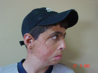
Fig. 2a,b Pseudotorticollis
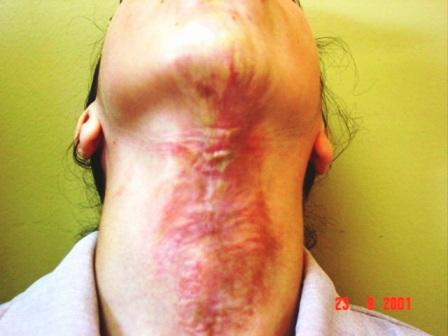
Fig.3a. Central neck scar (Preop.)
Fig.3b. Central neck scar (Postop)
Fig. 4a. Cheek scar (Preop)
Fig. 4b. 1 wk Postop
Fig.5a. Extensive face scar (Preop)
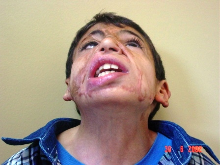
Fig.5b. Extensive face scar (Postop)
Having unburned skin adjacent to the area to be reconstructed facilitates efforts to improve the quality and function of the burn scar. The color, texture, thickness, elasticity, overall composition, and amount of hair of available donor skin need to be comparable to the desired (normal) qualities of the skin in the area to be reconstructed.(6)
Sir Harold Gillies has taught us that aesthetic surgery attempts to improve upon normal, whereas reconstructive surgery strives to restore the abnormal toward normal. It is difficult to separate these two agendas in burn reconstruction because form and function are so intimately intertwined.(6)
According to the modified principles from Feldman(13) and Neale et al.,(15) basic tenets of reconstruction in the neck are applied as follows: 1- delay reconstruction until scars have matured; 2- avoid creating vertical linear scars; 3- orient scars parallel to relaxed skin tension lines; 4- release extrinsic contracture before intrinsic contracture; 5- match donor skin according to thickness, color, and texture; 6- advance unburned skin caudally rather than cephalically when possible to decrease the possibility of iatrogenic lower lip ectropion; 7- reconstruct the contracture regions with the head and neck in full extension, rotation, and lateral flexion to minimize the postoperative tension; 8- resurface according to regional aesthetic units; 9- create the cervicomandibular contour; and 10- replace the contracture area with like tissue.
Reconstructive methods used after excision of post burn scar defects often focus on coverage of the defect without adequate attention to the types of tissues and methods used. Excision and direct closure of a scar is theoretically a wonderful option in that there is no donor site or skin quality problem. Unfortunately, this is rarely possible on the face or extremities without undue tension.(6)
Serial excision may be a useful technique in attempting to improve wide scars on the scalp, trunk and extremities. However, serial excision is generally unsatisfactory on the face because it either distorts features or results in unacceptably wide scars.(6) Although thin split-thickness skin grafts may be necessary acutely, they are rarely satisfactory for most definitive resurfacing of the face and scalp. Full-thickness skin grafts and composite grafts generally have better skin quality than thin split grafts owing to their dermal content.
A disadvantage of large full-thickness grafts is that they may require split grafts to close the donor sites. Also, when skin grafts are used, multiple procedures are often required to achieve the final result.(16)
Local or regional flaps fulfill all of the criteria for ideal skin quality where enough unscarred skin is available. Large cervico-pectoral transposition flaps can be used to resurface significant portions of the face with high-quality skin.(14) The biggest drawback to these multistage procedures is the unsightly split-thickness skin graft required to close the donor site.
Tissue expansion has afforded tremendous advances in burn reconstruction. It has all of the advantages of local or regional flaps, providing high-quality skin and minimal donor site deformity. However, there are disadvantages of this technique. At minimum, two surgical procedures are required. There is a considerable complication rate (25% to 48%) with risk of infection and exposure. It is not infrequent that underlying bone or cartilage demonstrates distortion and overlying fat may atrophy, and these effects may be permanent. It is cumbersome for the doctor and patient, requiring weekly injections.(8,15)
A more detailed prospective study investigating such matters as amount of exact scar excised, amount of advancement achieved at each stage of advancement, improvement in relevant neck angles and objective patient satisfaction scores is required. This will be conducted in the near future on a more harmonious group of patients now that we have a better understanding of the limitations of the procedure.
Conclusion
Many different techniques are available for reconstruction of a particular defect. The choice of technique depends on many factors including: Experience of surgeon, age and fitness of patient, willingness of both surgeon and patient to undergo the best possible reconstructive technique.
We found this technique useful in the late reconstruction of a large number of patients with mild to moderate facial and neck burns. It is easy, quick, repeatable and convenient to the patient with a low frequency of complications. It is an additional technique to the armamentarium of the reconstructive surgeon to deal with such difficult burns. It has limitations and is not suitable for the more extensive neck burn.
References
1.Gókalan L, Ozgúr F. Mavili E., Gúrsu G. Reconstruction of post burn face deformities. Annals of Burn and Fire Disasters 1997 Jun; volume X-n.2.
2.Jonsson CE, Dalsgaard CJ. Early excision and skin grafting of selected burns of the face and neck. Plast Reconstr Surg 1991; 88(1):83-92.
3.Cole JK, Engrav LH, Heimbach DM, et al. Early excision and grafting of face and neck burns in patients over 20 years. Plast Reconstr Surg 2002; 109:1266-1273.
4.Iwuagwu FC,Wilson D, Bailie F. The use of skin grafts in postburn contracture release, a 10-year review. Plast Reconstr Surg 1999; 103:1198-1204.
5.Tsai FC, Mardini S, Chen DJ, et al. The classification and treatment algorithm for post-burn cervical contractures reconstructed with free flaps. Burns, 2006; 32: 626-633.
6.Gottlibe LJ, Beahm EK.. Pediatric Burn Reconstruction. In: Michael L. Bentz, Pediatric Plastic Surgery, Vol. 2. Appelton & Lange, 1999. P619-633.
7.Woo SH, Seul JH. Preexpanded arterialized venous free flaps for burn contracture of the cervicofacial region. British J of Plastic Surgery, 2001; 54: 390-395.
8.Hudson DA, Grobbelaar AO. The use of tissue expansion in children with burns of the head and neck. Burns, 1995; 21(3): 209-211.
9.Lopez CE., Ferro A. Primary reconstruction of anterior neck burns with free flaps. British J of Plastic Surgery, 2005; 58: 102-105.
10.Kenney JG, DiMercurio S, Angel M. Tissue expanded radial forearm free flap in neck burn contracture. J. Burn Care & Rehabil.1990; 11(5): 443-445.
11.Jia CY, Chen B, Su Y. Prefabricated lined axial flaps for reconstruction of extensive post-burn facial and forehead full thickness composite defects. Burns 2002; 28: 688-690.
12.Hyakusoku H, Pennington DG, Gao JH. Microvascular augmentation of the super-thin occipitocervico- dorsal flap. Br J Plast Surg 1994; 47: 465-469.
13.Feldman JJ. Reconstruction of the burned face in children. In D. Serafin and NG. Georgiade (Eds.), Pediatric Plastic Surgery, Vol. 1. St Louis: Mosby, 1984.
14.Motamed S, Kalantar AJ, Marzban S. Expanded occipito-cervico-pectoral flap for reconstruction of burned cervical contracture. Burns 2003; 29: 842-844.
15.Neale HW, Kurtzman, LC, Goh KBC et al. Tissue expanders in the lower face and anterior neck in pediatric burn patients: Limitations and pitfalls. Plast Reconstr Surg 1993; 91(4): 624-631.
16.Feng-Chou T, Jui-yung Y, Mardini S, et al. Free Split-Cutaneous Perforator Flaps Procured Using a Three-Dimensional Harvest Technique for the Reconstruction of Postburn Contracture Defects. Plast Reconstr Surg 2004; 113(1):185-193.