Abstract
Objective:
To describe the demographics, clinical features, course, therapeutic interventions,
and outcomes of patients with Orf infection seen at the Royal Medical Services
Hospitals.
Methods: We
describe 64 patients with Orf infection who presented to dermatology clinics at
different Royal Medical Services Hospitals during a five year period from March
2002 to February 2007. Their clinical presentations, complications and treatment
were reported through regular follow-up at weekly intervals.
Results: Sixty
four cases (42 males and 22 females) were studied. Their ages ranged from 8-67
years. History of exposure to sheep or lambs was documented in 57 cases (89%).
The mean incubation period was seven days (ranged 4-15 days). The incidence
peaked after the feast of sacrifice each year. Thirty-six patients (56%) were
not aware about the infection before they had been examined by the
dermatologist. Misdiagnosis by physicians unacquainted with the disease led to
incision of the lesion in 21 patients (33%). The most common sites were fingers,
dorsum of hand, and palms. Diagnosis was made by history, appearance and
location of the lesion and clinical course.
In three cases, histopathological examination was made. The average
resolution time of the lesions was 32 days without significant scarring. Complications were reported in 21 patients
(33%). In all cases, symptomatic treatment and local wound care were applied.
Additionally, systemic antibiotics were used in 13 cases and cryotherapy in 11
cases.
Conclusion: Orf is an endemic infection in Jordan. Although
it is a self-limiting disease, prompt diagnosis is of paramount importance in
order to alleviate the anxiety of patients because the lesions could resemble other
more serious infections, and to avoid inappropriate treatments and possible complications.
A national emphasis on the cognizance of the infection, public awareness and
preventive measures is highly recommended.
Key
words: Ecthyma contagiosum, Orf, Royal
Medical Services
JRMS
December 2010; 17(4): 41-46
Introduction
Orf, also known as ecthyma contagiosum or contagious
pustular dermatitis, is a self-limiting zoonotic skin infection caused by an
epitheliotropic deoxyribonucleic acid (DNA) parapoxvirus.(1) It is a worldwide disease but little is known
about its prevalence and specific geographical distribution.(2)
Orf infection is endemic in sheep and goats and transmission to humans usually
occurs by direct contact with an infected or recently vaccinated animal or,
less often, indirectly through handling contaminated meat or objects, such as
fences, barn doors, feeding troughs, and shears in conjunction with skin trauma.(3,4)
The infection advances through six clinicopathological
stages and usually heals uneventfully in 4-6 weeks. The skin lesions are
commonly solitary and typically affect the hands and arms.(4,5) In humans, lasting immunity is
conferred by infection.(4,6)
Table
I. Summary of study findings
|
Sex
|
Males
- 42 (66%)
|
Females
- 22 (34%)
|
|
Age
|
Mean
- 39 years
|
Range:
8-67 years
|
|
Incubation
period
|
Mean
- 7 days
|
Range:
4-15 days
|
|
History
of exposure
|
Yes
- 57 (89%)
|
No
- 7 (11%)
|
|
Occupation
|
Farm
workers and shepherds (22)
Nonprofessional
slaughters (11)
|
Housewives
(15)
Butchers
(9)
Not
known (7)
|
|
Attending physician at
first presentation
|
Dermatology
clinics (32)
Surgeons
(16)
|
General
practitioners (7)
Emergency
room (3)
Orthopedic
surgeons (6)
|
|
Referral
diagnosis in 32 cases
|
Orf
infection in 11 (31%)
|
Incorrect
in 21 (69%)
|
|
Patient
awareness about infection
|
Yes
- 28 (44%)
|
No
- 36 (56%)
|
|
Initial
management
|
Incision
/excision in 21 (33%)
|
Antibiotics
in 24 (38%)
|
|
Laterality
|
Right
- 43 (67%)
|
Left
- 17 (27%)
Bilateral
- 4 (6%)
|
|
Number
of lesions
|
Solitary
- 38 (59%)
|
More
than one - 26 (41%)
|
|
Distribution
of 108 lesions
|
Fingers
(62)
Dorsum
of hand (23)
Aalm
(17)
|
Forearm
(3)
Arm
(1)
Leg
(1)
Face
(1)
|
|
Resolution
time
|
Mean
- 32 days
|
Range:
22-54 days
|
|
Complications
|
Yes
- 21 (33%)
|
No
- 45 (67%)
|
Table
II. Complications reported by 21
patients out of 64 patients with Orf infection
|
Complication
|
Number
of patients (%)
|
|
Regional
lymphadenopathy
|
12
(18.8)
|
|
Lymphangitis
|
8
(12.5)
|
|
Secondary
bacterial infection
|
7
(10.9)
|
|
Erythema
multiforme
|
5
(7.8)
|
|
Disseminated
papulo-vesicular rash
|
4
(6.3)
|
|
Scarring
|
2
(3.1)
|
|
Stiffness
of the digit
|
1
(1.6)
|
However,
some authors claim that immunity to Orf is not permanent as re-infection may
occur.(7,8) Skin lesions are commonly misdiagnosed and
patients overtreated. Notably, there is a scarcity of large-scale studies
addressing this disease in the literature.
Herein, we report the clinical features and management
of 64 patients with Orf infection providing insight into associated
complications and recent diagnostic and therapeutic concepts. To the best of
our knowledge, this is the first study reflecting the prevalence of this
endemic disease in Jordan.
Methods
We describe 64 patients with Orf infection who were
seen in different dermatology clinics at Royal Medical Services’ Hospitals
during a five year period from March 2002 to February 2007. The diagnosis of Orf
infection was mostly made by history, appearance and location of the lesion and
clinical course. Histopathological examination was only used to verify the
diagnosis in atypical cases. The data were analyzed with regards to patients’
sex, age and occupation, the clinical presentation, the diagnoses made by the
attending physicians, history of exposure, the incubation period (interval
between exposure and appearance of lesions), number and distribution of
lesions. Therapeutic modalities used in each case were also noted. Clinical
course, including the resolution time of the lesions, and complications were
reported through regular follow-up visits at weekly intervals.
Results
Sixty four patients (42 males and 22 females) with Orf
infection were studied. Their ages ranged from 8-67 years. Table I details
the demographic data and the clinical features seen in the study group. History
of exposure was documented in 57 cases (89%). The mean incubation period was seven
days (ranged 4-15 days). The incidence peaked in spring and after the feast of
sacrifice each year. The average duration of the disease before the patient
sought medical care was 13 days. Only 32 cases attended the dermatology clinics
directly, and 36 patients (56%) were not aware about the infection before they
had been examined by the dermatologist. Unfortunately, in most cases it was not
possible to verify whether the infection was from known infected animals or
not. Misdiagnosis by physicians unacquainted with the disease led to incision
of the lesion in 21 (33%) patients before referral. One hundred and eight Orf
lesions were encountered in 64 cases. The lesions were solitary in 38 cases
(59%), they were distributed mostly over upper limbs, and they affected the
right side in 43 (67%) cases. Pseudo-koebnerization was observed in three
patients. The diagnosis was made by history, appearance and location of the
lesion and clinical course. In three cases, histopathological examination was
made. The average resolution time of the lesions was 32 days (ranged 22-54 days)
without significant scarring. Complications, notably observed in the target and
acute stages, were reported in 21 patients (33%) (Table II). In all cases,
symptomatic treatment and local wound care were applied. Additionally, systemic
antibiotics were used in 13 cases and cryotherapy in 11 patients.
Discussion
Orf infection has been recognized as a common
occupational disease among those in contact with infected animals or
contaminated meat, such as farmers, butchers and veterinarians.(1,9)
The natural disease in sheep and goats usually occurs in the spring.(4)
It is characterized by proliferative papulo-vesicular lesions of the gums,
lips, nose, udder and groin, which usually resolve in 1-2 months.(2,10,11)
Infection transmission to human commonly
occurs after trivial trauma. Although rare, human-to-human spread and auto-inoculation
may also occur.(12)
Seasonal variation in the incidence of human Orf infection
has been previously noted(7) and was seen in our series. The
peak was noted during the spring time. However, Orf is not uncommon among
Muslims because many cases are seen after the feast of sacrifice(12,13)
and we observed an epidemic outbreak of human Orf after the feast of sacrifice
each year in Jordan.
After an incubation period of 3-8 days, the lesions
progress through six clinical pathological stages, each lasting about one week.(1,2,4) In the first ‘‘maculopapular’’ stage, an
erythematous macule becomes a papule. A lesion with a red centre, a white
middle ring and a red periphery characterizes the second ‘‘target’’ stage (Fig.
1). The third ‘‘nodular’’ stage consists of a reddish weeping nodule. A dry
lesion with small black dots on the outer surface develops in the fourth
‘‘regenerative’’ stage. In the fifth ‘‘papillomatous’’ stage, papillomas appear
on the lesion’s surface. A dry, thick crust is the main feature of the last
‘‘regressive’’ stage. Finally, the skin lesion usually resolves within 4-6
weeks with no residual scarring.
History of exposure was documented in 57 cases (89%).
The remaining seven patients may have acquired infection by indirect
contacts. Most patients (59%) had solitary lesions, and all of them had
incubation periods less than 15 days. Men were affected more than women because
they are more likely to be in direct contact with farm animals and
slaughtering. Most females were housewives and were involved in milking or
preparing the contaminated meat (especially during cleaning the animals’ heads)
before consumption.
Lesions occurred more on the right side and generally
on the dominant hand. The hands and arms are the body sites most frequently
affected by this virus.(4) This is consistent with the
findings in our sample. With two exceptions, all lesions occurred on the upper
extremities in our study. The extensor surface of index and thumb were the most
common affected sites. A facial lesion over the right cheek was observed in one
patient (Fig. 2). Previously, facial lesions have occasionally been reported.(2,4,5,14) Additionally, a solitary lesion was observed
over the left leg. Interestingly, linear pseudo-koebnerization on the
palm and dorsum of hand was observed in three patients overlying inadvertently
cut wounds (Fig. 3). These lesions appeared within one week after slaughtering
and evolved in the classical stages of Orf infection. This observation mostly
represents an auto-inoculation of the virus in this phenomenon.
Fifty-six percent of patients were not aware of the
infection before they had been examined by the dermatologist. Three patients
presented seeking medical advice for the associated complications but were not
aware of the Orf lesion. These
observations highlight the lack of public awareness about the infection.
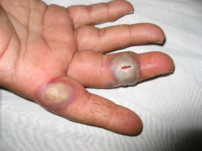
Fig. 1.
Two typical Orf lesions in the target stage. The one over the middle finger has
been incised by the surgeon
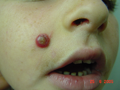
Fig. 2.
Facial Orf: erythematous weeping nodule, initially mistaken for Spitz nevus
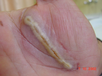
Fig.
3. Linear pseudo-koebnerization in
Orf infection overlying cut wound over the palm
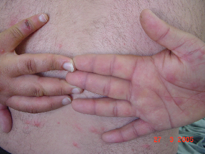
Fig.
4. Solitary Orf lesion over the left
little finger complicated by erythema multiforme over the palms and
disseminated papulo-vesicular eruption
Although Orf infection is a self-limiting disease,prompt
diagnosis is of paramount importance as it alleviates the anxiety of affected
patients and avoids inappropriate treatments and complications. Patients’
anxiety results from Orf lesions resembling other life threatening infections such
as cutaneous anthrax, tularemia and erysipeloid, and the fear of malignancy in
such a rapidly growing lesion. Skin lesions were commonly misdiagnosed as
pyogenic granulomas and pyogenic infections and patients were overtreated by
unacquainted physicians. This led to incision or excision of lesions in 33% of
patients before referral. We believe that late presentations and misdiagnoses
are likely attributed to the under-recognition of the disease.
The diagnosis is usually clinical, based on the
history of contact with infected animals, typical skin lesions and follow up.(15)
A biopsy can be done for histopathological examination if the diagnosis is in
question. The biopsy is best taken from an early lesion; showing hyperkeratosis
and cell vacuolization in the upper epidermis with eosinophilic
intracytoplasmic inclusions. However, the histological examination taken from our
patients showed features consistent with those seen in late Orf lesions.
Viral cultures, fluorescent antibody tests, electron
microscopy have all contributed to establish the diagnosis, but these are
rarely required and restricted to specialized centers.(2,16)
Milker’s nodule, a parapox viral infection transmitted from cows, cannot be
differentiated from Orf by its clinical appearance or by electron microscopy.(17) Only polymerase chain reaction
assay can definitively identify a parapoxvirus as Orf virus,(11,18)
but this is not yet routinely available.
In our study, all patients were immunocompetent. The
course of the disease was uneventful in the majority of patients. Additionally,
no recurrences have been observed in our patients. The reported complications
in our study are summarized in Table II; including erythema multiforme (EM) and
disseminated papulo-vesicular rash (Fig. 4).
Noteworthy, the incidence of complications was clinically
evident during the second and third stages and was much higher in the group who
received surgical intervention. Furthermore,
there was a
notable
delay of healing of the lesions in patients who developed superimposed
infections, EM or in whom the lesion was incised. A review of the literature on
human Orf infection yields a diverse array of reported sequelae. Most commonly,
fever, lymphangitis, lymphadenopathy, and secondary bacterial infection have
been noted.(19) Additionally, there have been some reported
cases complicated by EM, bullous pemphigoid, Stevens-Johnson syndrome, widespread
maculopapular or blistering eruptions, toxic erythema, and eyelid edema, as
well as giant, persistent or recurrent lesions in immunocompromised patients.(12,17,20-22)
Recently, a distinct and unique Orf-induced
immunobullous disease has been reported in two cases.(17)
Orf is a self limiting viral infection, which usually
regresses spontaneously in 6-8 weeks. Therefore, treatment of Orf is usually
symptomatic.(12) Surgery
can cause complications and thus must be avoided as a treatment for typical
lesions.(23) To date, there is no available specific
antiviral treatment and no human vaccine has been produced for the Orf virus.
Various treatments have been anecdotally reported; cryosurgery,(24)
idoxuridine,(25) imiquimod,(12) cidofovir(26)
and interferon(27) have been reported to reduce the time to
healing or to clear persistent infection. The use of antibiotics should be
restricted to patients with suspected secondary bacterial infection.
Cryotherapy is considered beneficial in patients presenting with early
infection.
We believe that Orf infection is more common in Jordan
than seen in clinical practice. However, because of the benign nature of the
disease and familiarity with infection, many individuals, and particularly
those involved with sheep rearing, are apt not to seek professional advice.
Physicians, especially non-dermatologists, who have not encountered many Orf lesions
should be aware of this disease and consider it in the differential diagnosis
of hand lesions. Barrier precautions and proper hand hygiene by farmers and
butchers and isolation of infected animals are recommended preventive measures.(1,18)
Conclusion
Orf infection is
an endemic self-limiting infection in Jordan. Prompt diagnosis of this
disease is of paramount importance to alleviate the anxiety of patients, and to
avoid inappropriate treatments and complications. A national emphasis on the
cognizance of the infection, public awareness and prevention measures is highly
recommended.
References
1. Georgiades G, Katsarou A,
Dimitroglou K. Human Orf (Ecthyma
contagiosum). Journal of Hand Surgery (British and European Volume)
2005; 30B: 4: 409-411.
2. Bodnar MG, Miller OF, Tyler WB. Facial Orf. Journal of American Academy
of Dermatology 1999; 40: 815-817.
3. Chahidi
N, de Fontaine S, Lacotte B. Human Orf. British
Journal of Plastic Surgery 1993, 46:
532-534.
4. Leavell
UW Jr, McNamara MJ, Muelling R, et al. Orf. Report of 19 human cases with clinical and pathological
observations. JAMA 1968; 204: 657-664.
5. Revenga
F, Paricio JF, del Agua C, Merino FJ. Facial Orf. J Eur Acad Dermatol Venereol
2001; 15: 80-81.
6. Diven DG. An overview of
poxviruses. J Am Acad Dermatol. 2001; 44(1):1-16
7. Groves RW, Wilson-Jones E, MacDonald DM. Human Orf and milkers’ nodule: a clinicopathologic
study. J
Am Acad Dermatol 1991; 25: 706-711.
8. Robinson AJ, Petersen GV. Orf virus infection of workers in the meat industry. N
Z Med J 1983; 96: 81-85.
9. Newson IE, Cross F. Sore mouth in sheep transmissible to man. J Am Vet Med Assoc 1934; 84: 790-802.
10. McKeever
DJ, Jenkinson DM, Hutchison G, Reid HW. Studies of the pathogenesis of
Orf virus infection in sheep. J Comp Pathol 1988; 99:
317-328.
11. Kottaridi
C, Nomikou K, Lelli R, et al. Laboratory
diagnosis of contagious ecthyma: Comparison of different PCR protocols with
virus isolation in cell culture. Journal of Virological methods 2006;
134: 119-124.
12. Erbagci Z, Erbagci I, Almila
Tuncel A. Rapid improvement of human Orf
(ecthyma contagiosum) with topical imiquimod cream: report of four complicated
cases. J Dermatolog Treat 2005; 16: 353-356.
13. Uzel M, Sasmaz S, Bakaris S,
Cetinus, et al. A viral
infection of the hand commonly seen after the feast of sacrifice: human Orf (Orf
of the hand). Epidemiol Infect 2005; 133: 653-657.
14. Gurel
MS, Ozardali I, Bitiren M, et al. Giant Orf on the nose. European Journal of Dermatology 2002; 12:
183-185.
15. Inceoğlu F. Orf (ecthyma contagiosum): an occasional diagnostic
challenge. Plast Reconstr Surg
2000; 106: 733-734.
16. Gill
MJ, Arlette J, Buchan KA, Barber K. Human Orf. A
diagnostic consideration? Archives of Dermatology 1990, 126: 356-358.
17. Schmidt
E, Weissbrich B, Bröcker EB, et al. Orf followed by erythema multiforme. J Eur Acad Dermatol Venereol
2006; 20: 612-613.
18. Centers for Disease Control
and Prevention (CDC). Orf virus
infection in humans-New York, Illinois,
California, and Tennessee, 2004-2005. MMWR Morb Mortal
Wkly Rep 2006; 55: 65-68.
19. White KP, Zedek DC, White WL, et
al. Orf-induced immunobullous
disease: A distinct autoimmune blistering disorder. J Am Acad
Dermatol 2008; 58: 49-55.
20. Mourtada
I, Le Tourneur M, Chevrant-Breton J, Le Gall F. Human Orf and erythema
multiforme. Ann Dermatol Venereol 2000; 127: 397-399.
21. Macfarlane AW. Human Orf complicated
by bullous pemphigoid. Br J Dermatol 1997; 137(4):656-7.
22. Hunskaar S. Giant Orf in a
patient with chronic lymphocytic leukaemia. Br J Dermatol 1986; 114:
631-634.
23. Bacakoglu AK, Ozkan M, Ekin A. Stay away from surgery: ecthyma contagiosum. Handchir Mikrochir
Plast Chir 2001; 33: 283-286.
24. Ocampo Candiani J, González
Soto R, Welsh Lozano O. Orf nodule: treatment
with cryosurgery. J Am Acad Dermatol 1993; 29: 256-257.
25. Freeman G, Bron AJ,
Juel-Jensen B. Ocular infection with Orf
virus. Am J Ophthalmol 1984; 97: 601-604.
26. Geerinck K, Lukito G, Snoeck
R, et al. A case of human Orf
in an immunocompromised patient treated successfully with cidofovir cream. J
Med Virol 2001; 64: 543-549.
27. Tan ST, Blake GB, Chambers S. Recurrent Orf in an immunocompromised host. Br J
Plastic Surgery 1991; 44: 465-467.