Abstract
Objective: To assess the dimension and types of root trunk of mandibular and maxillary first
molars and their influence on the diagnosis and management of molars with
furcation involvement.
Methods: A total of 105 extracted first molars were used in
this study. Included teeth comprised 41 maxillary first molars, and 64
mandibular first molars. For
each tooth, the vertical dimensions of the root trunk and root length were
assessed with a micrometer caliber. The types of root trunk were classified
according to the ratio of root trunk height to root length into types A, B and
C. Types A, B and C are defined as root
trunks involving the cervical third or less, up to half of the length of the
root, greater than the apical half of the root respectively. The length of root trunk and the prevalence of different types of root
trunk in maxillary and mandibular molars were analyzed. The percentage of root
trunk to root length was also calculated.
Results: Root trunk types A, B and C accounted for 34.9%, 61.8%,
and 3.3% of maxillary molars; 62.5%, 37.5%, and 0% of mandibular molars
respectively. In maxillary molars, the prevalence of type-A was found to be
39.1% and 41.4% on the buccal and distal root trunks respectively, while less
than that on the mesial root trunk (24.4%); the greatest
prevalence of type B was found on mesial root
trunk
(75.6%) while type C was found only on the distal root trunk (9.8%).
In mandibular molars, the type-A was found on buccal root trunk and
lingual root trunk with a higher
prevalence (73.4%, and 51.6%) than type B (26.6% and 48.4%) while no root trunk
type C was found in lower molars. The
mean root trunk dimension for
maxillary molars was 4.9, 4.31, and 3.9mm for the mesial, distal and buccal
respectively, while for mandibular molars 3.7mm for the buccal and 4.3mm for
the lingual. It was also noted that as the mean root trunk increased, the mean
root length decreased.
Conclusion: Awareness
of root trunk type and dimension may help the practitioner in the diagnosis,
treatment plan, and prognosis of periodontally involved molars.
Key Words: First permanent molar,
Furcation involvement, Root trunk
JRMS
March 2011; 18(1): 45-51
Introduction
Diagnosis, treatment, and
prognosis of furcation involvement (FI) are still challenging problems in the
field of periodontal therapy. The unpredictable results of periodontal therapy
in furcation-involved teeth is due in part to the complexity of furcation
morphology, such as cervical
enamel projection,bifurcational ridge, root proximity, length of root trunk
(RT), furcation entrance dimension, root fusion, and enamel pearls, for review
see Matthews and Tabesh.(1) Of these factors predisposing to
periodontal disease, enamel pearls were the only local anatomical factors
investigated in the Jordanian population, and were reported to have a
prevalence of 4.76%.(2)
The practical application of the anatomical knowledge
to clinical dentistry is mandatory to improve the overall dental health service
provided either as a preventive or therapeutic measure.
RT can be defined as the part of the root complex that
extends between the cemento-enamel junction and the furcation entrance. Its
height may be measured in millimeters or given in relation to the maximum
length of the root complex.(3) Hou and Tsai in 1997 developed a
classification scheme that takes into account the length of the RT compared to
total root length. Type A has the shortest RT involving a third or less of the
cervical area of the root, type B includes up to half of the length of the
root, while in type C the furcation entrance is in the cervical two-third of
the root or greater than the apical half of the root.(4)
The height of the RT in addition to the amount of
horizontal and vertical bone loss were suggested to supplement furcation
classification in order to facilitate the diagnosis, prognosis and treatment
planning.(5,6) Tunnel
preparation as a part of resective furcation therapy requires a short RT and a
wide diameter of the furcation entrance for proper postoperative plaque control
management by the patient.(7)
In regenerative therapy, RT dimension is considered one of the relevant
anatomical factors that may relate to the outcome of therapy.(8)
Furcation morphology of multi-rooted teeth, in
particular first molars has been investigated in the literature.(9,10)
However, The decision for a specific treatment mode for a
periodontitis-affected furcation certainly depends on a careful diagnosis.
Novel treatment modalities compel the therapist to acquire the necessary data
and to correctly interpret the respective observations. A thorough and detailed
diagnosis of all aspects of furcation involvement is demanding in clinical
experimentation.(3)
The objective of this study was to assess the
dimension and type of RT of mandibular and maxillary first molars and analyze their influence on the
diagnosis and management of molars with furcation involvement.
Methods
For the purpose of this study,
mandibular and maxillary molars were selected from an extracted teeth
collection of a dental practice disposal
of three Hospitals (Prince Aysha Medical Complex, Prince Rashed
Ben Al-Hassan
Hospital, and the
Out-patient Clinics of King Hussein Medical Center). Teeth selection was based firstly on having
intact roots and furcation regions, secondly on preserved cemento-enamel
junction unaltered by loss of tooth substance due to dental caries, fractures,
or tooth wear and thirdly on presence of
sufficiently intact crowns to facilitate sorting of teeth according to general anatomical
characteristics.
Of the 105 teeth retrieved, 41
were maxillary first molars, and 64 were mandibular first molars. To remove any
attached soft tissue, all teeth were immersed in 5.25% sodium hypochlorite for
30 minutes, and then sterilized by autoclave. If any calculus obscured the furcation entrances or the root trunk, this
calculus was removed gently using a manual curette scaler.
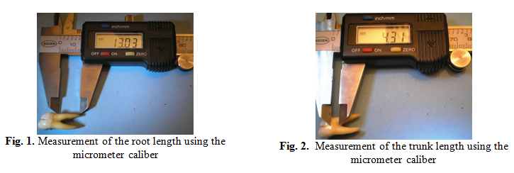
The vertical dimensions of the root trunk and root
length were assessed with a micrometer caliber (Fig 1, 2). Measurements of the
maxillary molars included the vertical height of the buccal root trunk (BRT),
mesial root trunk (MRT), distal root trunk (DRT), mesiobuccal root length
(MBRL), distobuccal root length (DBRL) and palatal root length (PRL). Similar measurements were obtained for mandibular molars, including buccal and lingual root trunks (BRT, LRT), mesial and distal root lengths (MRL DRL).
Table I. The mean ± SD and range for the root
and trunk lengths of the investigated teeth
|
Maxillary First Molar
(n:41)
|
Mandibular First Molar (no:64)
|
|
Mesiobuccal
RL*
|
Distobuccal RL
|
Palatal RL
|
Mesial RL
|
Distal RL
|
|
12.88±2.9 (9.9-16.7)
|
11.95±2.8
(9.1-15.01)
|
13.09±3.2 (10.01-17.3)
|
13.96±3.66 (9.98-20.01)
|
13.54±3.25
(9.97-20.12)
|
|
Mesial
TL**
|
Distal TL
|
Buccal TL
|
Buccal TL
|
Lingual TL
|
|
4.98±1.39
(3.2-7.7)
|
4.31±1.22
(3.4-7.8)
|
3.97±0.79
(2.8-7.6)
|
3.75±2.21
(2.4-6.5)
|
4.31±1.08
(2.6-6.6)
|
*RL: root length
**TL: trunk length
Table II. Types
of root trunk in relation to the length of root trunk and root length with the
percentage of the root trunk / root length in maxillary first molars
|
Maxillary
Molars
|
Type of
root trunk
|
Root n ( %)
|
Length of root trunk
mean±SD (mm)
|
Root length
Mean±SD
(mm)
|
% RT/RL
|
|
BRT
(41)
|
A
|
16 (39.1)
|
3.48±1.8
|
13.36±3.0
|
26.04
|
|
|
B
|
25 (60.9.)
|
4.99±1.2
|
12.14±1.6
|
41.1
|
|
|
C
|
0
|
0
|
0
|
|
|
MRT
(41)
|
A
|
10 (24.4)
|
3.63±0.9
|
14.4±2.0
|
25.2
|
|
|
B
|
31 (75.6)
|
5.23±0.8
|
12.73±2.5
|
41.08
|
|
|
C
|
0
|
0
|
0
|
|
|
DRT
(41)
|
A
|
17 (41.4)
|
3.48±1.12
|
13.18±1.17
|
26.4
|
|
|
B
|
20 (48.8)
|
4.99±0.1
|
12.32±1.5
|
40.5
|
|
|
C
|
4 (9.8)
|
6.1±1.5
|
10.87±2.12
|
56.11
|
|
Total
(123)
|
A
|
43 (34.9)
|
3.51±1.1
|
13.53±1
|
25.94
|
|
|
B
|
76 (61.8)
|
5.09±0.1
|
12.43±3
|
40.94
|
|
|
C
|
4 (3.3)
|
6.1±1.5
|
10.87±2.5
|
56.11
|
The types of root trunk were classified according to
Hou and Tasi (1997)(4) based on the ratio of root
trunk height to root length into types A, B and C. Types A, B and C are defined as root trunks
involving the cervical third or less, the cervical third to one half and greater
than the cervical half of the roots length, respectively. In order to determine
the type of root trunk, the mean root length for each aspect of the tooth was
measured e.g. the type of buccal root trunk of upper molars was
determined by measuring the mean length of buccal aspect roots which is the sum
of the mesiobuccal root length and distobuccal root length divided by two then
this measurement was correlated with the length of the buccal root trunk. The
length of root trunk and the prevalence of different types of root trunk in
maxillary and mandibular molars were analyzed.
Results
The range and mean values of root trunk and root
lengths for the examined teeth are presented in Table I. It can be perceived that, the shortest root dimension
of the maxillary molars was the distobuccal followed by the mesiobuccal then
the palatal (11.9, 12.8, 13mm). The
variation between the mean dimensions of the mesial (13.96) and distal roots
(13.54) of the mandibular first molars was less than 0.5mm. Regarding the root
trunk dimension, the buccal root trunk was the shortest in comparison with other
trunks for both molar types, the mean of the distal trunk of maxillary molars
and lingual trunk of mandibular molars were similar (4.31mm). The range of the
root trunk for maxillary molars was 2.4-6.6 mm, while for mandibular molars was
2.8-7.8 mm.
Table II and III
lists the ranges and mean values of the types of root trunk relative to the
dimensions of the root trunk and the root length in maxillary and mandibular
first molars respectively. In maxillary molars, the prevalence of type A was
found slightly greater on the DRT (41.4%) than the BRT (39.1%) while less than
that on the MRT (24.4%). The greatest prevalence of type B was found on MRT
(75.6%). In mandibular molars, type A
root trunk was found on BRT and LRT with a higher prevalence (73.4%, and 51.6%)
than type B (26.6% and 48.4%). No root trunks were classified as Type C except
for the distal root trunk of the maxillary molars, in which four trunks were
categorized as type C with a mean trunk length of 6.1mm and a mean root length
of 10.9mm. It can also be observed that
with increasing the mean root trunk there is a decreasing in the mean root
length. The above tables showed also the total distribution and prevalence of
root trunk types. Root trunk types A,B and C accounted for 34.9%, 61.8%, and 3.3% of maxillary molars; 62.5%, 37.5%, and 0% of mandibular molars respectively. It can be noted that the predominant root trunk for mandibular molars was type A while for maxillary molars was type B.
Table III. Types
of root trunk in relation to the length of root trunk and root length with the
percentage of the root trunk / root length for mandibular first molars
|
Mandibular
Molars
|
Type of
root trunk
|
Root n (%)
|
Length of root trunk
mean±SD (mm)
|
Root length
Mean±SD
(mm)
|
% RT/RL
|
|
BRT(64)
|
A
|
47 (73.4)
|
3.3±0.21
|
14.30±0.5
|
23.07
|
|
|
B
|
17 (26.6)
|
5±0.1
|
12.73±1
|
39.27
|
|
|
C
|
0
|
0
|
0
|
|
|
LRT(64)
|
A
|
33 (51.6)
|
3.7±2
|
14.57±3.92
|
25.39
|
|
|
B
|
31 (48.4)
|
4.83±0.1
|
12.67±0
|
38.12
|
|
|
C
|
0
|
0
|
0
|
|
|
Total
(128)
|
A
|
80 (62.5)
|
3.46±0.2
|
14.42±1.1
|
23.99
|
|
|
B
|
48 (37.5)
|
4.89±0.35
|
12.69±1
|
38.53
|
|
|
C
|
0
|
0
|
0
|
|
Table
IV. Comparison of root
trunk dimension of mandibular and maxillary molars between this study and other
studies
|
Author / year of publication
|
Maxillary Molars
|
Mandibular
Molars
|
|
MRT
|
DRT
|
BRT
|
LRT
|
BRT
|
|
|
4.98
|
4.31
|
3.97
|
4.31
|
3.75
|
|
Roussa 1998(13)
|
3.49
|
4.14
|
3.46
|
3.5
|
2.8
|
|
Plagmann et al. 2000(15)
|
4.8
|
4.5
|
4.3
|
4.3
|
3.3
|
|
Gher & Dunlap 1985(16)
|
3.6
|
4.8
|
4.2
|
|
|
Dunlap and Gher 1985(17)
|
|
4.0
|
4.0
|
|
Rosenberg 1988(18)
|
5.0
|
3.5
|
3.0
|
|
|
Mandelaris et al. 1998(19)
|
|
4.17
|
3.14
|
|
Kerns et al. 1999(20)
|
4.7
|
4.7
|
4.1
|
4.3
|
3.3
|
|
Porciúncula et al. 2007(21)
|
4.44
|
4.26
|
3.50
|
|
|
The percentage
of root trunk to root length was also calculated (Table II and III). For
maxillary first molars, it was found to be approximately 26%, 41% and 56% in
case of root trunk type A, B, and C respectively, while for mandibular molars,
this percentage was 24% and 38.5% for
type A and type B root trunk correspondingly.
Discussion
There has been a significant increase in the knowledge
and understanding of the etiology, pathogenesis, and treatment of inflammatory
periodontal diseases over the past few decades. However, arriving at a
diagnosis and determining the course of treatment are still based largely on
basic clinical and radiographic techniques, such as conventional assessment of
attachment and bone loss, which both have limitations. Therefore, knowledge of
the anatomical and morphological features of roots is necessary to achieve
better clinical practice in the field of periodontology. Extracted teeth is the
most commonly used method to measure the morphological features of teeth, as this
method provide a simple three dimensional insight profile view using different
angles. In addition, accurate measurements and re-measurements are easy to
perform and re-check at any point in time during the study or even afterwards.
Whilst handling of extracted teeth requires heat-sterilization prior to use for educational or
research purposes
according to infection control recommendations,(11) it was stated that autoclaving teeth does not appear to alter their physical properties or
dimensions.(12) For infection control purposes, teeth used in
this study were immersed in 5.25% sodium hypochlorite for 30 minutes followed
by autoclave sterilization prior to handling.
Root length is directly related to the quantity of
attachment supporting the tooth. Knowledge of root length is a critical element
that allows an informed clinical decision regarding diagnosis, prognosis, and
choice of treatment option of furcationally involved molars. The mean root
length for maxillary and mandibular first molars in the present study was 12.6mm
and 13.7mm respectively. For maxillary first molars the mean lengths of the
mesiobuccal and palatal roots were closer (12.9, 13mm) and longer than the
distobuccal root (11.9mm), while for mandibular first molars, the means of the mesial
and distal roots were 14 and 13.5mm respectively. Different results were
obtained by Roussa(13) for the maxillary molars who found
that the distobuccal is the longest root (12.2mm) compared to 11.3mm and 11.2mm
for the mesiobuccal and palatal respectively, while comparable results for
mandibular molars in which they found that the means for the mesial and distal
roots were 14.2 and 14mm respectively. However, racial variations in tooth morphology
are known to exist, particularly with respect to first permanent molars.(14)
The results of this study
regarding the mean root trunk height data for both maxillary and mandibular
first molars appears within the range of the same measurements in comparison
with other studies as shown in Table III.(13,15-21) Both
maxillary and mandibular first molars in this investigation had shorter root
trunks on the buccal aspects than on
lingual, mesial and distal sides, whereby mandibular first molars generally had
shorter root trunks than that of maxillary first molars, a finding that is also
comparable with others.(13,15,18-21) The length of
the mesial and distal root trunks of the maxillary first molar varies between
different studies. In the present study, the mean of the mesial root trunk length
was greater than the distal, this finding was in agreement with others(15,18,21)
while disagrees with some others who found that the distal root trunk was
longer than the mesial one.(13,16) Whereas a
similar root trunk length for the mesial and distal trunks was found by some
studies.(20)
Examination of proximal furcation is more difficult
than the buccal and lingual ones in particular when neighboring teeth are
present. This is often more difficult in case of long root trunks. Therefore,
such teeth may not be identified as furcationally involved without surgical
exposure. (20) A detailed knowledge of the length of the
variable root trunks in such sites is a fundamental prerequisite for the proper
interpretation of clinical data.
This investigation expressed the root trunk not only
as an amount measured by millimeters, but also as different types (A, B, and C)
and percentages according to its relation with the root length. This
information may help the practitioner to evaluate the amount and percentage of attachment
loss apical to the cemento-enamel junction required to expose the furcation for
the purpose of diagnosis in order to be able to establish a proper treatment
plan.
The maximum height of root
trunk for mandibular and maxillary molars in the present study was the 6.6mm,
and 7.8mm respectively. Dunlap and Gher(17) in their study on maxillary
first molars found no tooth had a root trunk longer than 6.0 mm. Hue et
al. found a significantly higher missing rate, poorer prognosis, and
inferior response to periodontal therapy for teeth with a long root trunk
length (type C). However, in this study
the overall percentage of root trunk type C was 3.3% for the maxillary molars
while none among the mandibular molars. Others found a greater percentage of
root trunk type C which comprises 11.9% in maxillary first molar and 1% in
mandibular first molars.(4)
The measurement of the
different types of root trunk relative to the dimensions of both roots and
trunks revealed that with increasing root trunk there is a decrease in root
length. This finding may influence the treatments choices of furcation involved
molars and the determination of
treatment plan for the furcationally involved molar. A furcation-involved molar with a long root
trunk and short roots may not be a candidate for root resection, since these
teeth lose more periodontal support with furcal invasion.(6) Horwitz et al.(23)
concluded that a long root trunk, a
wide furcation entrance and a furcation fornix coronal to the alveolar crest
have negative influences on the success of periodontal therapy.
The length and type of the
root trunk is one of the key anatomical factors that make molars particularly
susceptible to periodontal disease.(24) Mcclain and Schallhorn reported that short
root trunks surely influence the pathogenesis of furca involvement, a molar
with a short root trunk is more vulnerable to furcal involvement, but has a
better prognosis after treatment since less periodontal destruction has
presumably occurred.(8)
The results of the present study found that, in mandibular molars,
the minimum root trunk length at the buccal and lingual aspects was 2mm, so the
furcation could be approached even at the 2 mm probing attachment level. This
in turn leads to horizontal attachment loss and more progressive furcation
involvement. According to Dannewitz et al. molars with grade III FI had
the highest mortality and leads to a significant deterioration of prognosis.(25) On the other hand, molars with short
root trunks and more divergent roots have a more favorable prognosis when root
resective therapy is used.(26) A short root trunk and a wide furcation entrance
diameter are prerequisites for the indication of the tunnel preparation
procedure as a part of resective furcation therapy for the purpose of proper
postoperative plaque control management by the patient.(6) To ensure accessibility of the tunnel to plaque
control measures after tunnel preparation, the root trunk should reasonably
enough not be longer than a third of the total root length, i.e.,
approximately 4 mm based on figures by Paolantonio et al.(27)
However the variation in the
lengths of the root trunk between the buccal and lingual sides may interfere
with the treatment by tunnel preparation. In the present investigation we also
found that lingual root trunks of mandibular molars are on the average longer
than the buccal root trunks which might, after tunnel preparation, impede
accessibility for plaque control on the lingual furcation entrance.
The length and type of root trunk
are not the only factors that need to be considered in treatment planning in
case of furcation involvement. The presence of developmental grooves and concavities
on the trunk surface(28) is another factor that may
contribute to the outcome of regenerative periodontal therapy in case of short
root trunk. Lu(29) reported that 94% of the furcations
possessed variant depth of developmental concavities on the root trunks. These
superficial irregularities at the entrances of furcations may prevent complete
adaptation of the coronal microstructure of the membrane along their root
surfaces, so they suggested that subgingival application of guided tissue
membranes 1-2 mm below CEJ cannot ensure complete adaptation of furcation
defects with their coronal microstructures in the majority of molars. Kerns et
al. found that the mean CEJ to root groove distances ranged from 1.35 to
1.65 mm for maxillary first molars, and from 1.16 to 1.22 mm for mandibular
first molars.(20) Therefore regenerative periodontal
therapy in case of short root trunk could be compromised especially if
developmental concavities and grooves present on the root trunk.
The small number of teeth used in
this study can be considered as a limitation. Most of the first molars
extracted in our department(s) are badly destructed without a sufficiently
intact crown and root structure to facilitate sorting and measurement.
Limitations
of the Study
Further prospective studies to compare the effect of
different periodontal treatment modalities on molars with different types of
root trunk is needed.
Conclusion
Awareness of root trunk type and dimension may help
the practitioner in the diagnosis, treatment plan, and prognosis of
periodontally involved molars.
References
1. Matthews DC, Tabesh M. Detection of localized tooth related factors that predispose to periodontal infections. Periodontol
2004; 34: 136-150
2.Darwazeh
A, Hamasha AA. Radiographic evidence of enamel pearls in Jordanian dental
patients. Oral Surg
Oral Med Oral Pathol Oral Radiol Endod 2000 Feb; 89(2):255-258
3.Müller H-P, Eger T. Furcation diagnosis. J Clin Periodontol 1999; 26: 485–498.
4. Hou GL, Tsai CC. Types and dimensions of root trunk correlating with diagnosis of molar
furcation invohements. J Clin Periodontol 1997: 24: 129-135.
5.Hou
GL, Chen YM, Tsai CC, Weisgold
AS. A new classification of molar furcation
involvement based on the root trunk and horizontal and vertical bone loss.
Int J Periodontics Restorative Dent 1998; 18:257-265
6.Al-Shammari KF, Kazor CE, Wang H-L. Molar root anatomy and management of furcation defects. J Clin Periodontol 2001; 28: 730–740
7.Rüdiger
SG. Mandibular and maxillary furcation tunnel preparations – literature
review and a case report. J Clin Periodontol 2001; 28: 1–8
8.Mcclain
PK, Schallhorn RG. Focus on furcation defects – guided tissue
regeneration in combination with bone grafting. Periodontology
2000; (22): 190–212.
9. Hou
GL, Hung CC, Tsai CC, Weisgold
AS. Topographic study of root trunk type on
Chinese molars with Class III furcation involvements: molar type and furcation
site. Int J Periodontics Restorative Dent 2005 Apr;25(2):173-179
10.Santana RB,
Uzel MI, Gusman H, Gunaydin Y,
Jones JA, Leone CW.
Morphometric analysis of the furcation anatomy of mandibular molars. J Periodontol 2004 Jun;75(6):824-9
11.Kohn WG, Collins AS, Cleveland JL, et
al.
Guidelines for infection control in dental healthcare setting-2003; 52: 47
12.Parsell DE, Stewart BM, Barker JR, Nick TG, Karns L, Johnson RB. The
effect of steam sterilization on the physical properties and perceived cutting
characteristics of extracted teeth. J Dent Educ 1998; 62: 60–63.
13.Roussa, E. Anatomic characteristics of the
furcation and root surfaces of molar teeth and their significance in the
clinical management of marginal periodontitis. Clinical Anatomy 1998; 11:177–186.
14.Chiu BM, Zee KY, Corbet EF, Holmgren CJ. Periodontal implications of furcation entrance dimensions in chinese
first permanent molars. Journal of Periodontol 1991 May; 308-311
15.Plagmann H-C, Holtorf S,
Kocher T. A study on the imaging of
complex furcation forms in upper and lower molars. J Clin Periodontol
2000; 27: 926–931.
16.Gher MW, Dunlap RW. Linear variation of the root surface area of the maxillary
first molar. Journal of Periodontology 1985; 56:
39–43.
17.Dunlap
RM, Gher ME.
Root surface measurements of the mandibular first molar. Journal of
Periodontology 1985; 234-238.
18.Rosenberg MM. Furcation
involvement: periodontic, endodontic and restorative interrelationships. In:
Rosenberg MM, Kay HB, Keough BE, Holt RL, ed. Periodontal and prosthetic
management for advanced cases. Chicago:
Quintessence Publishing Co., 1988; 249-251.
19.Mandelaris GA, Wang HL, MacNeil RL. A
morphometric analysis of the furcation region of mandibular molars. Compendium of Continuing Dental Education 1998; 19: 113–120.
20.
Kerns DG, Greenwell H, Wittwer JW, et al. Root trunk
dimension of 5 different tooth types. Int J Periodontics Restorative Dent 1999
Feb; 19(1):82-91
21. Porciúncula HF, Zuza EP, da Porciúncula MM, de Toledo BE, Mendes AJ. Root trunk height as a risk factor for periodontal
furcation involvement in maxillary first molars: an in vitro study. J Int Acad Periodontol 2007 Jul; 9(3):89-95
22.Carnevale G, Pontoriero R, and Lindhe J. Treatment
of Furcation-Involved Teeth. In Lindhe J, Karring T, and Lang NP. Clinical
Periodontology and Implant Dentistry 4thed by Blackwell Munksgaard,
2003; 711.
23.Horwitz J, Machtei EE,
Reitmeir P, Holle R, Kim TS, Eickholz P.
Radiographic parameters as prognostic indicators for healing of class II
furcation defects. J Clin Periodontol 2004; 31: 105–111.
24.DeSanctis M, Murphy KG. The role of resective periodontal surgery in the treatment of furcation defects. Periodontology 2000.
2000; 22: 154–168
25.Dannewitz B, Krieger JK,
Hu¨sing J, Eickholz P. Loss of molars
in periodontally treated patients: a retrospective analysis five years or more
after active periodontal treatment. J Clin Periodontol 2006; 33: 53–61
26.Hempton T, Leone C. A Review of root resective therapy as a treatment
option for maxillary molars. JADA
1997; 128: 449-455
27.Paolantonio M, di Placido, G,
Scarano, A, Piattelli, A. Molar root furcation: morphometric and morphologic
analysis. International Journal of Periodontics and Restorative Dentistry
1998; 18: 489– 501.
28.Svdrdstrom G, Wennstrom JL. Furcation topography of the maxillary and mandibular
first molars. J Clin Periodontol 1988; 15: 271-275.
29.Lu HKJ. Topographical characteristics of root trunk
length related to guided tissue regeneration.
Journal of Periodontology 1992 Mar; 63: 215-219.