ABSTRACT
Objective:
The aim of this study is to assess the efficacy of Miconazole Oral Gel in the treatment of denture - induced stomatitis and to compare it to the use of
corrective and hygienic measures only.
Method:
Forty two prosthodontic patients, wearing removable prosthesis and presenting with denture stomatitis, were divided equally and randomly into two
groups. Group A received Miconazole Oral Gel in addition to oral hygiene instructions, denture cleansers, and correction of denture faults, and group B
received the same treatment with the exception of Miconazole Oral Gel.
Clinical and mycological examinations using Sabourauds Dextrose Agar Media were
done for each patient in both groups, and were repeated for each in the follow up visits. Data were collected, tabulated, and statistical analysis was
done to find inter- and intra-group differences.
Results:
Results revealed that candidal count and the erythema of the underlying mucosa were significantly reduced in both groups at follow-up visits compared
with the pretreatment condition, but the reduction was significantly more in the group that received Miconazole Oral Gel.
Conclusions:
Oral hygiene instructions play an important role in reducing severity of the condition, but they should be used in combination with Miconazole Oral Gel
to gain the best results.
Key words:
Candidal count, Denture-induced stomatitis, Miconazole Oral Gel.
JRMS December 2014; 21(4): 39-45 / DOI:
10.12816/0008064
Introduction
The human oral cavity contains a variety of species of bacteria and fungi, which can sometimes be the cause of different kinds of inflammation. (1) The most common inflammatory changes of mucosa seen under the denture are named stomatitis prosthetic, denture sore
mouth or denture stomatitis.(2)
Candida albicans
, the most commonly isolated opportunistic human fungal pathogen, can cause different skin and mucosal infections as well as life-threatening systemic
infections.(3,4) The involvement of C. albicans as a potential causative agent in denture-induced stomatitis was extensively
described, and it remains the most frequently isolated yeast in the oral cavity, nevertheless, other species have been isolated and are involved in
various diseases.(5) C. albicans associated stomatitis is a common disease that occurs in 50–60% of denture wearers. (6-8)
|
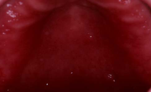
Fig. 1a:
Type1: Pin point stomatitis
|
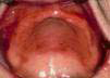
Fig. 1b:
Type2: diffuse denture stomatitis
|
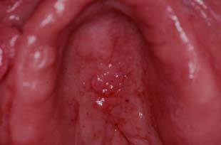
Fig. 1c:
Type3: Papillary stomatitis
|
|
Fig. 1.(a, b, and c)
Clinical Newton's classification of denture- induced stomatitis cases
|
Newton(9) had sorted this condition into three stages:
Stage 1: local or pin-point hyperaemia. (Fig. 1a)
Stage 2: diffuse hyperaemia. (Fig. 1b)
Stage 3: diffuse and hyperplastic. (Fig. 1c)
Denture stomatitis, caused mainly by C. albicans, can be observed in a high percentage of people wearing dental prosthesis. (10) C. albicans, which exist in the oral cavity as saprophyte, increase in number and gain pathogenicity after a prosthesis
is used.(11)
Attachment of C. albicans to dental prostheses is believed to be an important step in the development of denture stomatitis. Many factors are
involved in the adhesion of C. albicans to dental prosthesis like presence of sucrose in the medium, material characteristics like
surface roughness, surface hydrophobicity, electrostatic forces, composition of the material, type of matrix, size of fillers, and configuration of
fillers.(12-17)
However, the etiology of denture stomatitis appears to be multifactorial; its development is influenced by the presence of a dental prosthesis, C. albicans and other microorganisms, but also by other local and systemic factors such as an acid salivary pH, high carbohydrates ingestion,
long-term antibiotic therapy, hormonal therapy as well as systemic illnesses such as diabetes mellitus or arterial hypertension.(19)
Treatment of denture stomatitis involves three measures: correction of the denture, oral hygiene, and antifungal drugs, although many practitioners try
to keep off medications in general and antifungal medications in these cases to avoid their elderly patients any extra cost or health burden.
Therefore, this research was done to evaluate the benefit of adding antifungal medication specifically Miconazole Oral Gel to treat a relatively poor
socioeconomic class and medically impaired group of patients.
Methods
This study was performed at Princess Haya Al-Hussein Hospital and Prince Ali Hospital after obtaining ethical approval from the Royal Medical Services
Human Research Ethics Committee.
Our sample was selected from the patients that attended the Prosthodontic clinic complaining from signs and symptoms of denture stomatitis. The sample
was collected over the period from the 3rd of January, 2012 to the 26th of April, 2012.
The original sample consisted of 50 patients, eight were excluded because of missed follow up visits. The final study sample consisted of 42 Jordanian
patients (24 (57.1%)) females and 18 ((42.9%) males) ranging in age from 33 to 85 years, wearing heat-curing acrylic removable prosthesis, presenting
with denture stomatitis, and reported wearing their dentures for more than 12 hours a day.
Exclusion criteria included: smoking, alcoholism, diabetes,
autoimmune diseases, immunosuppression, and drugs. The observers were two trained prosthodontists who examined the patients and directly observed the
swabbing and inoculation procedures in the lab.
The patients were informed about the study course and they were asked to sign a written consent. Patients were divided equally and using simple
randomization into two groups regardless of their gender. Those in group A, with the mean age was 63.7 ±5.8 years, were treated with Miconazole Oral
Gel in addition to correction of denture faults, oral hygiene instructions, and using hypochlorite denture cleanser once daily and normal saline mouth
wash three times daily. Instructions were given for all patients in group A
Table I:
Age and gender distribution of both groups A and B
|
Variable
|
Gender
|
n (%)
|
Age (Years)
|
|
Mean
|
S. D.
|
|
Group A
|
F
|
13 (31)
|
63.7
|
5.79
|
|
M
|
8 (19)
|
|
Group B
|
F
|
11 (26.2)
|
62.5
|
13.34
|
|
M
|
10 (23.8)
|
|
Total
|
42 (100)
|
63.1
|
10.178
|
Table II:
Clinical findings and dental status at presentation among patients of the whole sample
|
|
Group A
|
Group B
|
|
Clinical Newton's stage
|
Stage I
|
Stage II
|
stage III
|
Total
|
Stage I
|
Stage II
|
stage III
|
Total
|
|
Number (%)
|
6(28.6)
|
12(57.1)
|
3(14.3)
|
21(100)
|
7(33.3)
|
12(57.1)
|
2(9.5)
|
21(100)
|
|
Dental status
|
|
|
Edentulous
|
18
|
21
|
14
|
21
|
|
Partially dentate
|
1
|
3
|
|
Combination
|
2
|
4
|
|
Prostheses
|
|
|
CU*/CL**
|
19
|
21
|
15
|
21
|
|
PU^/PL^^
|
0
|
2
|
|
Combination
|
2
|
4
|
|
Oral /denture hygiene
|
|
|
Good
|
0
|
0
|
0
|
0
|
0
|
0
|
0
|
0
|
|
Moderate
|
4
|
3
|
0
|
7
|
5
|
0
|
0
|
5
|
|
Poor
|
2
|
9
|
3
|
14
|
1
|
12
|
2
|
16
|
*CU complete upper, **CL complete lower, ^PU partial upper, ^^PL partial lower to use the Miconazole Oral Gel on the palatal mucosa and on the denture base twice daily.
Those in group B, with mean age 62.5 ±13.3 years, were treated with corrective measures only (all the previously mentioned means but without
Miconazole). Mean ages and gender distribution of both groups are summarized in Table I.
Clinical Examination
Personal data of the patients including name, gender, age, occupation, and social status was collected. A detailed medical and dental history was
obtained from each subject in both groups. Intra-oral examination included assessment of the dental status, oral and denture hygiene (according to the
degree of cleanness of the prosthesis), and a detailed clinical examination of the oral mucosa.
The erythematous lesions in the palatal mucosa in the sample were grouped according to the clinical appearance into stages I, II or III, according to Newton’s classification.
The dentures in all subjects were evaluated for their retention, stability, maxillomandibular relationships, and occlusion. Clinical examinations were
performed by prosthodontists. The clinical examination and Newton's staging were done at presentation visit, after two weeks, and after four weeks, for
both groups and are shown in Table II. Examination of the palatal mucosa clinically revealed that 13 cases (31%) were presenting in Newton’s stage I,
24 cases (57.1%) were in stage II, and five cases (11.9%) were in stage III.
At presentation, 30 patients (71.4%) had poor oral/denture hygiene and 12 patients (28.6%) had moderate oral/denture hygiene. Prosthesis examination of
the whole sample was investigated and found to have 34 patients (81%) were completely edentulous patients, and two patients (4.8%) were partially
dentate patients, and six cases (14.3%) were combined cases.
Table III:
The frequency of clinical and mycological scores in both groups
|
Group A
|
|
Newton Staging
|
Clinical Scores
|
Mycological scores
|
|
Start
|
2 wks
|
4 wks
|
Start
|
1 wk
|
2 wks
|
3 wks
|
4 wks
|
|
n (%)
|
n (%)
|
n (%)
|
n (%)
|
n (%)
|
n (%)
|
n (%)
|
n (%)
|
|
0
|
0(0)
|
13(61.9)
|
18(85.7)
|
0(0)
|
0(0)
|
8(38.1)
|
14(66.7)
|
18(85.7)
|
|
1
|
6(28.6)
|
5(23.8)
|
3(14.3)
|
6(28.6)
|
7(33.3)
|
8(38.1)
|
7(33.3)
|
3(14.3)
|
|
2
|
12(57.1)
|
3(14.3)
|
0(0)
|
10(47.6)
|
11(52.4)
|
5(23.8)
|
0(0)
|
0(0)
|
|
3
|
3(14.3)
|
0(0)
|
0(0)
|
4(19.0)
|
3(14.3)
|
0(0)
|
0(0)
|
0(0)
|
|
4
|
-
|
-
|
-
|
1(4.8)
|
0(0)
|
0(0)
|
0(0)
|
0(0)
|
|
Total
|
21(100)
|
21(100)
|
21(100)
|
21(100)
|
21(100)
|
21(100)
|
21(100)
|
21(100)
|
|
Group B
|
|
Newton Staging
|
Clinical Scores
|
Mycological scores
|
|
0
|
0(0)
|
1(4.8)
|
6(28.6)
|
0(0)
|
0(0)
|
1(4.8)
|
1(4.8)
|
1(4.8)
|
|
1
|
7(33.3)
|
11(52.4)
|
11(52.4)
|
7(33.3)
|
8(38.1)
|
11(52.4)
|
13(61.9)
|
14(66.7)
|
|
2
|
12(57.1)
|
7(33.3)
|
4(19.0)
|
10(47.6)
|
9(42.9)
|
8(38.1)
|
7(33.3)
|
6(28.6)
|
|
3
|
2(9.5)
|
2(9.5)
|
0(0)
|
3(14.3)
|
4(19.0)
|
1(4.8)
|
0(0)
|
0(0)
|
|
4
|
-
|
-
|
-
|
1(4.8)
|
0(0)
|
0(0)
|
0(0)
|
0(0)
|
|
Total
|
21(100)
|
21(100)
|
21(100)
|
21(100)
|
21(100)
|
21(100)
|
21(100)
|
21(100)
|
Table IV:
The mycological and clinical response to treatment in both groups
|
(Group A)
|
Mycological Scores Mean
|
Clinical Newton's Scores Mean
|
|
Mean
|
SD
|
S.E.M.
|
Mean
|
S.D.
|
S.E.M.
|
|
At presentation
|
2.00
|
.183
|
.837
|
1.57
|
.163
|
.746
|
|
One week
|
1.81
|
.148
|
.680
|
|
|
|
|
Two weeks
|
.86
|
.173
|
.793
|
|
|
|
|
Three weeks
|
.33
|
.105
|
.483
|
|
|
|
|
Four weeks
|
.14
|
.078
|
.359
|
.14
|
.078
|
.359
|
|
(Group B)
|
|
|
|
|
|
|
|
At presentation
|
1.90
|
.181
|
.831
|
1.52
|
.148
|
.680
|
|
One week
|
1.81
|
.164
|
.750
|
|
|
|
|
Two weeks
|
1.43
|
.148
|
.676
|
|
|
|
|
Three weeks
|
1.29
|
.122
|
.561
|
|
|
|
|
Four weeks
|
1.24
|
.118
|
.539
|
.90
|
.153
|
.700
|
Table V:
Inter and intra- group comparison regarding both of the mycological and clinical scores.
|
Group
|
Test type
|
Time Interval
|
Mean
|
S.D.
|
S.E.M.
|
t-value
|
P value
|
|
A
|
Mycological
|
Starting visit
|
2.00
|
.183
|
.837
|
13.000
|
.000
|
|
Four weeks
|
.14
|
.078
|
.359
|
|
Clinical
|
Starting visit
|
1.57
|
.163
|
.746
|
10.954
|
.000
|
|
Four weeks
|
.14
|
.078
|
.359
|
|
B
|
Mycological
|
Starting visit
|
1.90
|
.181
|
.831
|
5.292
|
.000
|
|
Four weeks
|
1.24
|
.118
|
.539
|
|
Clinical
|
Starting visit
|
1.52
|
.148
|
.680
|
5.701
|
.000
|
|
Four weeks
|
.90
|
.153
|
.700
|
|
A-B
|
Mycological
|
Starting visit
|
2.00
|
.183
|
.837
|
-.357
|
.724
|
|
1.90
|
.181
|
.831
|
|
Four weeks
|
.14
|
.078
|
.359
|
9.312
|
.000
|
|
1.24
|
.118
|
.539
|
|
Clinical
|
Starting visit
|
1.57
|
.163
|
.746
|
-.237
|
.815
|
|
1.52
|
.148
|
.680
|
|
Four weeks
|
.14
|
.078
|
.359
|
4.985
|
.000
|
|
.90
|
.153
|
.700
|
Laboratory investigation
Swabs were used to collect samples from the fitting denture surface and the underlying mucosa from the rugae area, and were directly inoculated on Sabourauds Dextrose Agar Culture Medium, and were then incubated at 37°C for two days as shown in Fig. 2.

Fig. 2:
Colonies of Candida were counted after its inoculation in Sabouraubs Dextrose Agar plate at 37 C° for two days
Colony counts were made according to the following scale to obtain the yeast score: 0 (no growth), 1 (low growth), 2 (medium growth), 3 (high growth),
and 4 (confluent growth).
Study Course
Each patient in both groups was seen after one, two, three, and four weeks. Laboratory examination was done at these time intervals. While the clinical
examination and Newton's staging were performed at two and four week intervals.
Data were collected, tabulated, and proper statistical analysis were done. Paired Sample Student t-test was used to compare inter and intra- group mean
differences. Statistical Package for the Social Sciences (SPSS) software, version 16 was used in this study.
Results
The erythema of the underlying mucosa was significantly improved in both groups at follow-up visits compared with the pretreatment condition. Data of
Newton's staging at presentation, at two, and at four week intervals were compared for both groups as shown in Table III. Amounts of yeast colonies
were calculated for both groups A and B at each visit and were summarized in Table III. The mycological and clinical score means were analyzed, and
summarized in Table IV for both groups.
The number of colonies was reduced more significantly in group A than in group B as shown in Table IV. During follow up, the patients in group A, who
were using Miconazole Oral Gel, were noticed to have a significant improvement. At the end of treatment the percentage of patients showing complete
clinical cure was 85.7%.
The Percentage of patients showing complete mycological cure was 85.7% (Table III). While for group B, after four weeks
of oral hygiene instruction (cleaning the denture and using normal saline mouth wash) and corrective measures (correction of base and occlusion
errors), there was a significant reduction in yeast count in 66.7% of patients, while there was no change in yeast count in 33.3% of cases. Complete
cure was difficult to achieve in group B, as only one case (4.8%) was recorded to have negative mycological result (Table III).
There were no significant differences between both groups at the presentation visit in both the mycological and the clinical tests (p>0.05). While,
after four weeks there was reduction in C. albicans count in both groups, but for group A, there was a more significant reduction than in
group B (Table V).
Discussion
The results of this study revealed that there was a direct proportional relation between C. albicans colonies count found in the mycological
test and the Newton staging for the same case. This result was similar to other results of studies which concluded that there is a significant relation
between Newton staging of stomatitis and amount of yeast colonies.(20-23)
In some patients of the sample in this study, C. albicans showed to be resistant and was still exhibited even in low numbers in group A in 14.3%
of patients or in low to moderate numbers in 95.3% in group B patients. This scene has been studied by many authors.(7)
Results of this study showed that, the relationship between denture cleanliness and denture stomatitis is well established. Denture cleanliness did not
cure denture stomatitis completely, as evidenced by both the clinical and the microbiological scores.
Inter-group differences showed highly significant decrease in C. albicans counts in group A more than in group B (p<0.01), this indicates
that educating patients to improve their denture hygiene without providing adjunctive antifungal therapy is an impractical denture stomatitis
treatment. The C. albicans count of dentures in group B was greater than in the group A at all review visits.
In this study oral hygiene and the corrective measures of the prosthesis showed a significant reduction in number of yeast colonies, but gave no cure
for the lesions. On the other side, using Miconazole Oral Gel, in group A, induced a more significant reduction in numbers and gave cure in 85.7% of
the patients. This result was very close to the result of Dias et al.(25) who found that, Miconazole Oral Gel is an efficient
drug of choice for treatment of denture stomatitis.
The overall results of this study revealed that, Miconazole Oral Gel oral gel could be a beneficial drug for the management of denture
stomatitis, on the other hand, oral hygiene instructions and corrective measures may reduce yeast count to a lesser extent. These results concerning
the efficacy of Miconazole Oral Gel in treatment of denture stomatitis were consistent with the results of other studies.(24-26)
Conclusions
Miconazole Oral Gel oral gel plays an important role in reducing the severity of denture stomatitis but it should be used in combination with oral
hygiene instructions and correcting any denture faults.
Recommendations:
First:
1. Correcting any denture faults.
2. Advising patients to avoid nocturnal denture wear.
3. Oral hygiene instructions.
Second:
Instructing the patient to use Miconazole Oral Gel oral gel twice daily, by painting.
References
1. Aas JA, Paster BJ, Stokes LN, et al. Defi ning the normal bacterial flora of the oral cavity. J Clin Microbiol
2005; 43:5721-5732.
2. konsberg R, Axell T. Treatment of Candida-infected denture stomatitis with a Miconazole Oral Gel lacquer. Oral Surg OralMed Oral Pathol 1994: 78: 306-311.
3. Ihmels J, Bergmann S, Berman J, et al. Comparative gene expression analysis by differential clustering approach: application to
the Candida albicans transcription program. PLoS Genet 2005; 1:e39.
4. Jones T, Federspiel NA, Chibana H, et al. The diploid genome sequence of candida albicans. Proc Natl Acad Sci USA 2004: 101:
7329-7334.
5. Ramage G, Tomsett K, Wickers BL, et al. Denture stomatitis: a role for Candida biofilms.Oral Surgery, Oral Medicine, Oral Pathology, Oral Radiology and Endodontology 2004
; 98(1): 53-59.
6. Budtz-Jorgensen E. Ecology of Candida-associated denture stomatitis. Microb Ecol Health Dis 2000; 12:170-185.
7. Darwazeh AMG, Al-Refai S, Al- Mojawel S. Isolation of Candida species from the oral cavity and fingertips of complete denture wearers. J Proshet Dent 2001: 86: 420-423.
8. Pıres FR, Santos EBD, Bonan PRF, et al. Denture stomatitis andsalivary Candida in Brazilian edentulous patients. J Oral Rehabil 2002; 29:1115-1119
9. Newton AV. Denture sore mouth. A possible aetiology. Br Dent J 1962: 112:357-360
10. Baena-Monroy T, Moreno-Maldonado V, Franco-Martínez F, et al. Candida albicans, Staphylococcus
aureus and Streptococcus mutans olonization in patients wearing dental prosthesis. Med Oral Patol Oral Cir Bucal 2005;10:E27-E39
11. Abaci O, Haliki-Uztan A, Ozturk B, et al. Determining Candida spp. Incidence in Denture Wearers. Mycopathologia 2010: 10.1007/s11046-010-9275-8
12. San Millan
R, Ezkurra PA, Quindós G, et al.
Effect of monoclonal antibodies directed against Candida albicans cell wall antigens on the adhesion of the fungus to polystyrene. Microbiology 1996; 142: 2271-227.
13. Cannon RD, Chaffin WL. Oral colonization by Candida albicans. Crit Rev Oral Biol Med 1999; 10: 359-383.
14. Radford DR, Challacombe SJ, Walter JD. Denture plaque and adherence of Candida albicans to denture-base materials in vivo and in
vitro. Crit Rev Oral Biol Med 1999: 10:99-116.
15. Holmes AR, Gopal PK, Jenkinson HF. Adherence of Candida albicans to a cell-surface polysaccharide receptor on Streptococcus gordonii. Infect Immun 1995; 63:1827-1834.
16. Willis AM, Coulter WA, Hayes JR, et al. Factors affecting the adhesion of Candida albicans to epithelial cells of insulin-using
diabetes mellitus patients. J Med Microbiol 2000; 49: 291-293.
17. Cerano-Graner C, Cerero-Lapiedra R, Campoo-Trapero J, et al. In Vitro Study: Adherence of Candida Albicans to Acrylic Resin
Relationship to Surface Energy. Int J Prosthodont 2005: 18:392-398.
18. Nevzatoglu EU , Özcan M, Kulak-Ozkan Y , et al . Adherence of Candida albicans to denture base acrylics and silicone-based
resilient liner materials with different surface finishes. Clin. Oral Investig 2007; 11: 231.
19. Sanchez-Vargas LO, Pérez-Ros P, Romo-Garca J, et al. Minacin de pH salival y cultivo en pacientes con candidiasis bucal. Rev Iberoam Micol 2002; 19: 155-160.
20. Mojon P. Oral health and respiratory ınfection. J Can Dent Assoc 2002; 68(6):340-345.
21. Mahmoudabadi AZ, Drucker DB, Mandall N, et al. The oral yeast flora: effect of upper removable orthodontic appliances. Microb Ecol Health Dis 2002; 14:149-152.
22. Pereıra-Cenci T, Delbelcury AA, Crielaard W, et al. Development of Candida-associated denture stomatitis: new insights. J Appl Oral Sci 2008; 16(2):86–94.
23. Bartie KL, Williams DW, Wilson JM, et al. PCR fingerprinting of Candida albicans associated with chronic hyperplastic
candidosis and other oral conditions. J Clin Microbiol. 2001;39:4066–75.
24. Niimi M, Firth NA, Cannon RD. Antifungal drug resistance of oral fungi. Odontology. 2010; 98:15-25.
25. Dias A., Samaranayake A., Lee M. Miconazole Oral Gel lacquer in the treatment of denture stomatitis: Clin Oral Invest 1997: 1: 47–52.
26. Kulak-Ozkan Y, Kazazoglu E, Arikan A Oral hygiene habits, denture cleanliness, presence of yeasts and stomatitis in elderly people. J Oral Rehabli 2002: 29; 300-304.