ABSTRACT
Congenital Hemidysplasia with Ichthyosiform nevus
and Limb Defects (CHILD) syndrome is a very rare syndrome. It is
inherited as X-linked dominant trait. The cutaneous lesions and
extracutaneous defects are peculiar; as they involve one half of the
body. We present here two Jordanian cases of CHILD syndrome, a new born
girl and her mother. Up to our knowledge these cases are the first
reported cases of CHILD syndrome in Jordan. The rarity of this syndrome
prompted us to report these two cases.
Key words: CHILD, Syndrome
JRMS June 2015; 22(2): 51-54 / DOI: 10.12816/0011369
Introduction
CHILD syndrome; a syndrome of Congenital Hemidysplasia with Ichthyosiform nevus and Limb Defects, is a rare multisystemic disorder, with only 60 reported cases in the literature.(1) While most reports described sporadic cases, only 5 cases of mother to daughter transmission have been documented.(2) The syndrome is inherited as an X-linked dominant trait which is lethal in male embryos.
It’s characterized by unilateral inflammatory epidermal nevus and ipsilateral limb hypoplasia. It’s caused by a defect in cholesterol synthesis as a result of mutation in NSDHL (NADAPH sterol dehydrogenase - like) gene. (3)
We’re to report two cases of CHILD syndrome, a Jordanian newborn female patient, and her mother. In both of them the right side of the body has been affected.
Case report
A 10-day old-girl, the first born child of a non-consanguineous marriage. A product of full term normal vaginal delivery, and uneventful antenatal history, her birth weight was 3 Kg. Her mother had no history of known miscarriages.
She presented with erythematous, scaly, and velvety thickening of the skin over the right half of her body since birth, and a deformity of her right leg.
Cutaneous examination revealed an inflammatory nevus involving the right half of the body, with midline demarcation, covering the right half of anterior thorax and abdomen, sparing the face and scalp, with yellow-brown, waxy scaling covering the affected side. There are also few streaks of involvement on the contralateral side.
Ptychotropism (verrucous scaly plaques, with marked preference for flexural areas), markedly involving the flexural creases of the axillae and groins. The right labia showed hypertrophy, with velvety and maceration of the skin. Her right foot is deformed and looks like as if she has one large toe with a pendulous heel. Her mother also has an inflammatory nevus affecting the right side of her body, with few streaks over the left side of the trunk following Blaschko’s line. She also has skeletal deformity of her right hand and foot. She has bowing of her right index finger and syndactyly of both right third and fourth toe. (Figs 1-4)
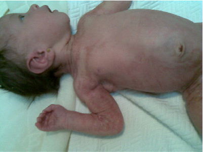

Fig. 1: Cutaneous lesions in a new born girl, showing erythematous,
scaly, and velvety thickening of the skin over the right half of the
body
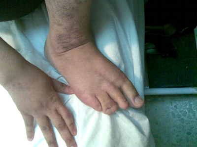
Fig.
4: Right upper and lower limbs deformity in a mother, bowing and
shortening of her right index finger and syndactyly of both right third
and fourth toe
According to the mother the grandmother also had similar lesions over her right lower limb. The grandmother gave birth for 3 daughters. Two of them were completely normal i.e. not having any cutaneous anomalies.
The newborn patient was thoroughly investigated to look for other associated congenital anomalies, but none was found. Complete Blood Count, Kidney Function and Liver Function tests all were within normal limit. Renal ultrasound was normal. 2D-cardiac echo showed normal pulmonary artery and aortic arch, and intact interatrial and ventricular septae. Skeletal survey concluded: no skeletal abnormalities, nor an evidence of chondrodysplasia puctata. And a chest X-ray was also normal.
Cutaneous lesions were managed conservatively using emollients and topical antibiotics for macerated area over the genitalia. She was evaluated by an orthopedic surgeon, who advised a below knee amputation, at the age of one year. A plastic surgeon was also consulted about vulvular lesion, and advised a conservative management.
The diagnosis of CHILD syndrome in these cases was done based on the well evident clinical picture, no genetic studies were done.
Discussion
The term CHILD syndrome was first introduced by Happle et al. (4) Though the earliest description of CHILD syndrome was reported by Otto Sachs in 1903. (5) It’s clinically characterized by unilateral erythematous hyperkeratotic verrucous scaly plaques, with marked preference for flexural areas (ptychotropism). (6) The ptychotropism was well evident clinically in the axillae and groins of the new born patient. Lesions are usually limited to one side of the body, usually the right side, with sparing of the face and oral mucosa.(7) Some patients had in addition to that a streaky distribution of erythematous patches following Blaschko’s line in the “unaffected” body side, with both linear and diffuse unilateral patterns coexisting in the same patient.(8) Both of our patients had right sided lesions with contralateral streaky blaschkoid patches; sparing the face, as what was mentioned in most reported cases. Konig et al reported bilateral almost symmetrical involvement in a woman with a novel mutation of NSDHL gene. (9) Associated with an Ipsilateral hypoplasia of the limbs, (4) most likely to affect long bones.(8) The severity of limb involvement varies from complete absence of the entire limb, as what was seen in the new born patient of having severely deformed foot, to absence or hypoplasia of small bones of the hands or feet, as what was seen in her mother. (So both the mother and her daughter present the two extreme of the disease regarding limb involvement). And other internal organ hypoplasia (e.g., brain, lung, thyroid, muscles, cranial nerves, heart, etc.). If a congenital cardiac defect is present in a patient (in left sided CHILD), it will be the main cause of early death. (4)
CHILD nevus can be distinguished from all other types of epidermal nevi by characteristic features such as ptychotropism, waxy yellowish scaling, a unique lateralization pattern showing both diffuse and linear involvement. (10, 11) This was well evident clinically in the skin lesions of both of our patients. So the clinical picture alone was enough for the diagnosis of CHILD syndrome. The peculiar feature in these two cases is that both the mother and her daughter had full picture of CHILD syndrome, the grandmother may have had partial manifestations of the syndrome. So recognition of these skin disorders
is important to decrease the risk of genetic transmission of the disease. Because a woman with an isolated CHILD nevus has an increased risk of giving birth to a daughter suffering from a complex congenital disorder, the CHILD syndrome. (10)
Mutations in the NSDHL (NADPH sterol dehydrogenase-like) gene cause CHILD syndrome. This gene is responsible for an enzyme production that is involved in cholesterol synthesis. Although high cholesterol levels are a well-known risk factor for heart disease, the body needs some cholesterol to develop and function normally both before and after birth. Cholesterol is an important component of cell membranes and the myelin sheath. Also, cholesterol plays an important role in the production of steroid hormones and digestive bile acids. (3) So low cholesterol levels disrupt the growth and development of many parts of the body. (3) It is not known, however, how a disturbance in cholesterol production leads to the specific lateralization of CHILD syndrome.
Treatment with topical emollients and retinoid can provide little improvement. (1) Recently surgical treatment with dermabrasion and skin grafting has been tried, yielding favorable outcome. (12)
Limitation of the study
No genetic study was done for these cases.
Conclusion
The CHILD syndrome is an inherited X-linked dominant, male-lethal trait. Recognition of this particular skin disorder is important for genetic counseling to decrease the risk of genetic transmission of the disease. Because a woman with an isolated CHILD nevus has an increased risk of giving birth to a daughter suffering from a complex congenital disorder, the CHILD syndrome .
References
1. Chander R, Varghese B, Jabeen M, et al. CHILD syndrome with thrombocytosis and congenital dislocation of hip: A case report from India. Dermatology Online Journal 2010; 16(8):6
2. Bittar M, Happle R, Grzeschik KH, et al. CHILD syndrome in 3 generations: the importance of mild or minimal skin lesions. Arch Dermatol 2006 Mar: 142(3): 348-351.
3. Caldas H, Herman G. NSDHL, an enzyme involved in cholesterol biosynthesis, traffics through the Golgi and accumulates on ER membranes and on the surface of lipid droplets. Hum Mol Genet 2003; 12(22): 2981-2991.
4. Happle R, Koch H, Lenz W. The CHILD syndrome: congenital hemidysplasia with ichthyosiform erythroderma and limb defects. Eur J Pediatr 1980; 134: 27-34.
5. Bittar M, Happle R. CHILD syndrome avant la letter. Journal of the American Academy of Dermatology 2004; 50(2): 34-37.
6. Happle R. Ptychotropism as a Cutaneous feature of the CHILD syndrome. J Am Dermatol 1990; 23:763-766.
7. Moulin G, Barrut D, France MP, et al. CHILD syndrome: naevus epidermique et hemidysplasia corporelle hypoplasique homolaterale. Ann Dermatol Venereal 1982; 109: 793-794.
8. Atherton GJ, Moss C. Rook's textbook of dermatology. 7th ed. Oxford: Nevi and other developmental defects. Wiley-Blackwell, 2004; pp; 15.21-15.22.
9. Konig A, Happle R, Fink-puches R, et al. A novel missense mutation of NSDHL in all unusual case of CHILD syndrome showing bilateral, almost symmetric involvement. J Am Acad Dermatol 2002; 46: 594-596.
10. Happle R, Mittag H, Kuster W. The CHILD nevus: a distinct skin disorder. Dermatology 1995; 191: 210-216.
11. Happle R. The group of epidermal nevus syndromes capsule summary. Journal of the American Academy of Dermatology 2010; 63(1):1-22.
12. Konig A, Skrzypek J, Loffler H, et al. Donor dominance cures CHILD nevus. Dermatology 2010; 220 (4): 340-345.