ABSTRACT
Objectives:
To evaluate the effect of oral propranolol in the treatment of infantile peri-orbital and/ or orbital capillary hemangioma.
Methods:
We conducted a prospective study at Prince Rashid Bin Al Hassan military hospital between 5th of August 2012 and 29th of January
2013. Eleven patients with peri-orbital and/or orbital capillary hemangiomas were included in this study.
All patients underwent complete ophthalmic
examination. Capillary hemangiomas were assessed, reporting their size, location, extension, and effect on the surrounding structures. Follow up
duration ranged from two to six months.
Results:
The age of patients with peri-orbital and/ or orbital capillary hemangiomas ranged from three to 17 months with a mean of 7 ± 4.92 months. Male: female
ratio was 1: 1.75. The upper eyelid was involved in seven (64%) cases, all of them had ptosis, three (43%) had dystopia, one (14%) had imbrication, and
one (14%) had squint.
The lower eyelid was involved in two (18%) cases; one of them was associated with lower lip capillary hemangioma. Orbital
involvement was seen in eight (73%) cases, six (75%) of them were associated with upper eyelid involvement and two (25%) with lower eyelid involvement.
Concurrent extra-ocular localization of hemangiomas was present in five (45%) cases (one in lip, one in tongue, 2 in forehead, and one in cheek).
Duration of treatment ranged between one and 30 weeks with an average of 14.2 ± 11.4 SD weeks. The dose of propranolol ranged between 1- 2 mg/kg with
an average of 1.2 mg/kg and only one patient with upper eyelid and forehead extension needed 4 mg/kg.
The color blanched in all patients after one
week. The capillary hemangioma decreased in size after one week of treatment in two (18%) patients, and in all of them after one month. Complete
regression of the hemangioma was seen in two (18%) cases one after two months and the other after 4 months of treatment. Five (45%) cases had
astigmatism before the start of treatment (mean ±SD, 0.9 ± 0.379 D) diopters and improved to mean ±SD, 0.56 ± 0.586 D.
Conclusion
: Oral propranol can be used as a modality for therapy of infantile capillary hemangioma.
Key words:
Infantile Capillary Hemangioma, Orbital, Peri-Ocular, Propranolol.
JRMS December 2014; 21(4): 53-60 / DOI:
10.12816/0008066
Introduction
Infantile capillary hemangiomas are soft tissue hamartomas that are predominantly seen in females below the age of one year,(1) and
about 10% of children usually develop one or more hemangiomas shortly after birth and before the end of first year of their life.(2)
Capillary hemangioma usually starts shortly after birth; enters a phase of rapid growth that lasts usually for few months, then stabilizes
for another few months, and involutes spontaneously in the majority of cases.(1,3) Although the natural course of
capillary hemangiomas is self- involution but treatment is needed when vital or sensory functions are impaired or disfigurement is anticipated. (2,4)
Capillary hemangioma is the commonest benign tumor of the peri-orbital and orbital area with a predilection for the
upper eyelid.(2,5) It may have deep extension into the orbit, and sometimes may extend superficially to involve the skin of the face.
Capillary hemangiomas are classified according to their location with respect to the skin and orbital septum into: cutaneous, purely preseptal,
preseptal with extraconal element, and combination of preseptal, extraconal and intraconal(6) Diagnosis is made clinically
but imaging studies such as ultrasound, CT scan, or MRI are usually needed for classification or diagnosis if it is deep in the orbit. (7) Periorbital and/ or orbital capillary hemangioma treatment is indicated if it causes visual impairment that may lead
to amblyopia due to ptosis, dystopia, astigmatism, anisometropia, or proptosis leading to exposure keratopathy, or optic nerve compression. (6)
Other indications include tumor necrosis, infection, and disfigurement.(8) Treatment is usually given in the
proliferative phase to prevent serious complications. Classically systemic steroid or intra-lesional steroid injections(1) were used
and in case of non-response to steroid, other modalities were used namely interferon,(9) chemotherapy,(4) topical
imiquamod,(10) laser,(11) or radiotherapy.(1)
These modalities of treatment are
associated with many complications and efforts were needed to look for a new safer modality. In 2008 Le’aute’-Labre’ze serendipitously observed
dramatic decrease in size of capillary hemangiomas in patients treated with propranolol,(12) which is a non- selective β-
adrenergic receptor blocker, for cardiac problems.
Methods
Complete ophthalmic examination was performed, including visual fixation preference, slit lamp and fundus examination, refraction under cycloplegia,
and ocular motility. Capillary hemangiomas were assessed, reporting their size, location, extension, and effect on the surrounding structures. Orbital CT scan was ordered
when necessary.
All patients were assessed by a pediatric senior specialist before the commencement of treatment. Assessment included clinical
examination, echocardiography, abdominal ultrasonography and CBC. Patient with associated oral or perioral hemangioma was examined by a pediatric
dental specialist. Exclusion criteria included patients with congestive cardiac failure, bronchial asthma, obstructive pulmonary disease, or patients on concurrent
treatment with other modalities.
Informed consent was signed by parents before commencement of treatment. Colored photographs were taken and the dimensions of the tumors were recorded
for all patients before treatment and in each follow up visit to monitor the response to treatment.
Patients were started on propranolol 1 mg/kg in the clinic and observed for six hours for vital signs. Mothers were then instructed to dissolve the
tablet and divide into doses followed by a bottle of milk. The dose was increased gradually by 0.2 mg/kg increment till the color changed and tumor
became soft. The patients were seen after two days, one week, and then monthly till the tumor disappeared or stabilized in size.
The main outcome measure was post treatment regression of lesion size and bleaching of its color. Secondary outcome measures included improvement in
astigmatism, amblyopia, and visual acuity, and safety of therapy. Simple statistics like mean, average, frequency, and percentages were calculated. We obtained approval of the Ethical Committee of the Royal Medical
Services.
Results
Eleven patients with peri-orbital and/ or orbital capillary haemangiomas were included in this study. The age ranged from three to 17 months with a
mean±SD of 7 ± 4.92 months. Male to female ratio was 1: 1.75. One child was born prematurely as one of a triplet.
|
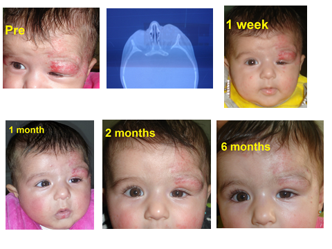
Fig. 1:
Left upper eyelid infantile capillary hemangioma with forehead and orbital extension. More than 50% decrease in size after six months
with improvement of dystopia and partial improvement of ptosis.
|
One patient had brain atrophy and esotropia. The upper eyelid was involved in seven (64%) cases, six (86%) in the left side and one (14%) in the right
side and all of them (100%) had ptosis, three (43%) had dystopia, one (14%) had imbrication, and one (14%) had convergent squint (stable 30Δ angle,
alternating in primary position with cross-fixation and refraction under cyclplegia was +1 diopter).
The lower eyelid was involved in two (18%) cases;
one of them was associated with lower lip capillary hemangioma. Orbital involvement was seen in eight (73%) cases, six (75%) of them were associated
with upper eyelid involvement and two (25%) with lower eyelid involvement (Fig. 1). Concurrent extra-ocular localization location of haemangiomas was
present in five (45%) cases (one in lip, one in tongue, two in forehead, and one in cheek).
One case had left forehead capillary hemangioma associated
with a large tongue hemangioma, which was partially excised, treated with radiotherapy and propranolol and recurred after drug was stopped. Duration of
treatment ranged between one and 30 weeks with an average of 14.2± 11.4 SD weeks.
The dose of propranolol ranged between 1- 2 mg/kg with an average of
1.2 mg/kg. Only one patient with upper eyelid and forehead extension needed 4 mg/kg (Fig. 2). The color blanched in all patients after one week. The
capillary hemangioma decreased in size after one week of treatment in two (18%) patients, and in all of them after one month. Complete regression of
the hemangioma was seen in two (18%) cases one after two months and another after four months of treatment (Fig. 3).
The average length of the lesion
before treatment was 3.2±1.6SD mm and dropped to 1.6±1.5SD mm. The average width was 2.5±1.4SD mm and decreased to 1.2±0.95SD mm. The average height,
measured with a ruler from the lateral side of the face and compared with other side, was 0.6±0.47 mm decreased to 0.28±0.43SD mm.
One patient had
recurrence of the hemangioma after recurrent vomiting due to gastrointestinal tract infection and the lesion was controlled and decreased in size after
restarting treatment. Two (18%) cases had regrowth of the hemangioma when propranolol was tapered over two weeks after almost complete involution and
stabilization for one month. The previous dose was re-started and the lesion decreased in size again.
Table I shows patients’ demographic data, characteristics of the capillary hemangiomas, indication of treatment, and concurrent extra-ocular capillary
hemangiomas.
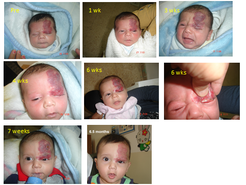
Fig. 2:
Left upper eye capillary hemangioma with forehead and orbital extension presented at the age of 3 weeks with severe ptosis and dystopia. Skin and
eyelid margin ulcerations started after 6 weeks of treatment and required a 4 mg/kg propranolol
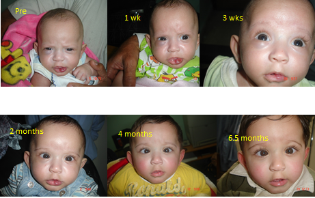
Fig. 3:
Complete involution of right lower eyelid and lower lip capillary haemangiomas after 4 months of treatment with 1.2 mg/kg propranolol in premature
infant. Esotropia started 2 months of treatment.
Table I:
Patients’ demographic data, characteristics of the capillary hemangiomas, indication of treatment, and concurrent extra-ocular capillary hemangiomas Five (45%) cases had astigmatism before the start of treatment (mean ±SD, 0.9 ± 0.379 D) diopters and improved to (mean ±SD, 0.56 ± 0.586 D).
|
No
|
G
|
Age
|
Size/Length
|
Size/Width
|
Height
|
Duration
(week)
|
Location
|
Orbit
|
Indication
|
|
Before
|
Final
|
Before
|
Final
|
Before
|
Final
|
|
1
|
F
|
7
|
2.5
|
0.5
|
2.5
|
0.5
|
0
|
0
|
28
|
UEL/ OS
|
+
|
Ptosis/ Med Dyst
|
|
2
|
F
|
3
|
2
|
1
|
1
|
0.5
|
0
|
0
|
4
|
UEL/ OS
|
-
|
Ptosis
|
|
3
|
M
|
3
|
3
|
0
|
2
|
0
|
0.5
|
0
|
26
|
LEL/OD
|
-
|
Up dyst/ Squint
|
|
4
|
F
|
10
|
3
|
1
|
3
|
1
|
1
|
0.1
|
30
|
Forehead
|
-
|
|
|
5
|
F
|
17
|
1
|
0.3
|
1
|
0.3
|
0.4
|
0.1
|
6
|
LEL/OD
|
+
|
|
|
6
|
F
|
9
|
3
|
2
|
2
|
2
|
0.5
|
0.1
|
10
|
UEL/ OS
|
+
|
ptosis
|
|
7
|
F
|
9
|
3
|
3
|
3
|
2
|
1
|
0.5
|
3
|
UEL/ OS
|
+
|
Ptosis/
dyst
|
|
8
|
M
|
12
|
3
|
1
|
2
|
0.5
|
0.5
|
0.3
|
24
|
UEL/OD
|
+
|
ptosis/
squint
|
|
9
|
F
|
3
|
7
|
1
|
6
|
1
|
1
|
0.3
|
20
|
UEL/OS
|
+
|
ptosis/
dystopia
|
|
10
|
M
|
1
|
5
|
5
|
3
|
3
|
0.2
|
0.2
|
4
|
UEL/OS
|
+
|
ptosis/
imbrication
|
|
11
|
M
|
3
|
2.5
|
2.5
|
2
|
2
|
1.5
|
1.5
|
1
|
LEL/OD
|
+
|
|
|
Avg.
|
M:F 4:7
|
7
|
3.18
|
1.57
|
2.5
|
1.163
|
0.6
|
0.282
|
14.18
|
UEL: 7
|
Orbit: 8
|
Ptosis:7
|
|
±SD
|
|
4.92
|
1.58
|
1.46
|
1.36
|
0.945
|
0.473
|
0.433
|
11.408
|
LEL:3
|
|
Globe Disp.: 4
|
|
|
|
|
|
|
|
|
|
|
|
|
|
squint:2
|
G: Gender, UEL: Upper eyelid, OS: Oculus Sinister, MedDyst: Medial Dystopia, LEL: Lower Eyelid, OD: Oculus Dexter, Up Dyst: Upward dystopia.
Table II:
Astigmatism before and after treatment.
|
Number
|
Pre
|
Post
|
|
1
|
-0.75 X 170
|
-0.25 X170
|
|
2
|
-1.00 X 180
|
-0.25 X 180
|
|
3
|
-0.5 X 170
|
00
|
|
4
|
-1.00 X 15
|
-1.50 X 15
|
|
5
|
-0.75 X 180
|
-0.5 X 180
|
|
Average ± SD
|
-0.9 ± 0.379
|
-0.56 ± 0.586
|
Table II
shows patients with astigmatism before and after treatment.
The patient with tongue hemangioma had good improvement in terms of eating, breathing, drooling and talking. None of the patients had propranolol side
effects. Follow up duration ranged from two to six months.
Discussion
Infantile capillary hemangioma is a self-limiting soft tissue hamartoma that involutes with time in the majority of cases with no serious sequelae.
Propranolol was found to inhibit the growth and enhances the involution of capillary hemangiomas by inducing vasoconstriction that leads to immediate
change in color and a palpable softening of the hemangiomas. This is believed to occur through down-regulation of the RAF-mitogen-activated (RAF,
Rapidly Accelerating Fibrosarcoma) protein kinase pathway that decreases the expression of VEGF (Vascular Endothelial Growth Factor) and bFGF (basic Fibroblast Growth Factor) genes and this explains the progressive improvement of the hemangioma, and the triggering of apoptosis of
capillary endothelial cells.(13)
Propranolol 10 mg tablet is very affordable; a 20/pack costs 1.2 JD.
Most of the studies in the literature were pilot studies with small samples.(12-24) There was no study conducted in Jordan on the
treatment of periocular hemangiomas with propranolol. By studying the effect of propranolol on infantile hemangioma in Jordan, we aim at encouraging
ophthalmologists and pediatricians to replace steroid therapy. In our study the average age was about seven months, which is almost the same as previous studies but higher than Fridman et al (24) where the average age was 3.1 months. This is probably as we focused on treating cases with risk of complications, the rate of
which is known to increase with age.
The female predominance in our study conforms with all previous studies in literature.(12-24) We included a child who was born
prematurely one of a triplet and had significant lower eyelid hemangioma at the age of three months. Because of risk of amblyopia this child was
started on oral propranolol 1mg/kg and monitored and followed up closely but, unfortunately, he developed esotropia with cross fixation and
insignificant refractive error.
The other two of triplets did not develop esotropia. Another patient who had cerebral palsy had also congenital
esotropia not related to the hemangioma or treatment.
The upper eyelid was most commonly affected site and most of them were on the left side, a finding to which we could not find an explanation. As all of
them had ptosis, and as some had dystopia, imbrication, or squint, treatment was indicated as soon as possible to decrease the risk of amblyopia. The first signs of improvement seen in our patients in terms of change in color and softening of the hemangiomas were seen within the first week, and
this is due to the vasoconstrictive effect of propranolol. These findings agree with previous similar studies.(12-14,17,20-24)
The last follow up showed a drop in surface area, size, and consistency of the hemangioma by almost 50%. This was lower than that of Missoiet al
,(25) and Sans et al,(13) probably due to lower average dose of treatment we used, 1.2 mg/kg in comparison
to 2 mg/kg in the former and 2-3mg/kg in the latter studies. Another explanation is the duration of the treatment which was short, 14.2 weeks, in our
study, 6.8 months and 6.1 months respectively and longer in Missoiet al (25) 6.8 months and in Sans et al (13) 6.1 months.
This reduction in size can be graded as good according to Haider et al,(26) who attempted to define the various grades of success as
follows: excellent results if the size of the hemangioma decreased by more than 50%, good if it decreased to 50%, fair if no further growth, and poor
if it continued to grow or the patients had intolerable side effects. The reduction in size of all lesions and the regrowth in two cases after tapering of propranolol and the reduction in size after giving the previous
dose give strong evidence of the effect of propranolol in the treatment of infantile capillary haemangioma.
Five patients had astigmatism of about 1 diopter before treatment that improved to 0.56 diopter after treatment, none of them had amblyopia based on
visual fixation preference tests. This is on contrary to Missoi et al,(25) who had 41% amblyopic children before treatment and
those with astigmatism of more than 1.5 diopters had improvement in astigmatism of 0.66 diopters (33%). All patients with concurrent extra-ocular location (45%) of hemangiomas had complete involution after treatment either simultaneous, before, or after
periocular hemangiomas.
The patient with tongue hemangioma was previously treated with surgery and radiotherapy because of risk of airway obstruction and given propranolol by
the oral surgeon. After the cessation of treatment the patient had regrowth of
the lesions and we started him on propranolol again and he improved in terms of eating, breathing, talking, drooling, decrease in size of the tongue,
and less tongue protrusion.(27)
None of the patients had significant propranolol side effects. None of them had hypoglycemia as the dose was given immediately before the meal. None had fatigue, hypotension. One patient had
gastroenteritis that was not related to propranolol. Bronchial asthma in terms of shortness of breath or wheezes was not noticed. Other side effect
such as sweating, cold hands, and bradycardia, diarrhea, and sleep disturbances were not reported.
Limitations of this study
The main limitations were the small sample size, and lack of control or comparative groups. This is due to infrequent patients with significant
periocular hemangiomas, and short follow up period. So a large prospective comparative study comparing the topical β- blocker,(28)
systemic and intra-lesional steroids, and oral propranolol is needed.
Conclusion
Oral propranolol can be used as a modality for therapy of infantile capillary hemangioma. However, a bigger contolled comparative case series would
give further proof and evidence.
References
1. Bang GM, Setabutr P.Periocular capillary hemangiomas: indications and options for treatment. Middle East Afr J Ophthalmol 2010;17(2):121-128.
2. Jalil A, Maino A, Bhojwani R, Vose M, et al. Clinical review of periorbital capillary hemangioma of infancy. J Pediatr Ophthalmol Strabismus 2011;48(4):218-225. Epub 2010 Jul 22.
3.
Alcántara-González J
, Boixeda P,
Truchuelo-Díez MT
, et al.
Infantile hemangiomastreated by sequential application of pulsed dye laser and Nd:YAG laser radiation: a retrospective study.
Actas Dermosifiliogr
2013; 104(6):504-511. Epub 2013 Mar 21.
4.
Donnelly LF
, Adams DM, Bisset GS 3rd
.
Vascular malformations and hemangiomas: a practical approach in a multidisciplinary clinic.
AJR Am J Roentgenol
2000; 174(3):597-608.
5. Nigwekar SP, Nigwekar PV. Atypical presentation of capillary hemangioma of upper eyelid: A case report. Pravara Med Rev 2011; 3(3).
6. Kanski JJ, Bowling B. Orbit. In: Clinical Ophthalmology: A Systematic Approach. London. Elsevier: 7th edition 2011; p.103-105.
7.
Li HY
, Xu YK,Lin BQ, Yu T.
Magnetic resonance imaging findings of capillary hemangioma in the brain. Nan Fang Yi Ke Da XueXueBao 2009; 29(5):1043-5, 1048.
8. Durairaj VD.
Treatment of deep orbital hemangiomas of infancy: an overview.
Arch Facial Plast Surg
2006; 8(3): 217-220.
9. Hastings MM, Milot J, Barsoum-Homsy M, Hershon L, et al.
Recombinant interferon alfa-2b in the treatment of vision-threatening capillary hemangiomas in childhood.
J AAPOS
1997; 1(4): 226-230.
10. Mascarenhas R, Guiote V, Agro J, Henrique M. Ulcerated infantile hemangioma treated with imiquimod. Dermatol Online J 2011;
17(9):13.
11. Brauer JA, Geronemus RG.
Laser treatment in the management of infantile hemangiomas and capillary vascular malformations.
Tech Vasc Interv Radiol
2013; 16(1):51-54.
12. Le´aute´-Labre`ze C, Dumas de la Roque E, Hubiche T, Boralevi F. Propranolol for severe haemangiomas of infancy. N Engl J Med 2008; 358:
2649–2651
13.
Sans V
, de la Roque ED,Berge J, Grenier N,
et al.
Propranolol for severe infantile hemangiomas: follow-up report. Pediatrics 2009; 124(3): e423-31.
Epub 2009 Aug 10.
14. Cruz OA, Siegfried EC. Propranolol treatment for periocular capillary haemangiomas. J AAPOS 2010; 3: 251-256.
15. Al Dhaybi R, Milet A, McCuaig C, Ospina L, Powell J. Treatment of periocular infantile haemangioma with propranolol: review of 17 cases. Pediatr Dermatol 2009; 26: 665-666.
16. Fay A, Nguyen J, Jakobiec FA, et al. Propranolol for isolated orbital infantile haemangioma. Arch Ophthalmol 2010; 128:
256-258.
17. Cheng JF, Gole GA, Sullivan TJ. Propranolol in the management of periorbital infantile haemangioma. Clin Experiment Ophthalmol 2010;
38:547-53.
18. Hogeling M, Adams S, Wargon O. A randomized controlled trial of propranolol for infantile hemangiomas. Pediatrics 2011; 128: 259-266.
19. Zvulunov A, McCuaig C, Frieden IJ, et al. Oral propranolol therapy for infantile hemangiomas beyond the proliferation phase: a
multicenter retrospective study. Pediatr Dermatol 2011; 28:94-98.
20. Celik A, Tiryaki S, Musayev A, et al. Propranolol as the first-line therapy for infantile hemangiomas: preliminary results of two
centers. J Drugs Dermatol 2012;11:808-811.
21. Mohanan S, Besra L, Chandrashekar L, Thappa DM. Excellent response of infantile hemangioma associated with PHACES syndrome to propranolol. Indian J Dermatol Venereol Leprol 2012; 78:114-115.
22. Chai Q, Chen WL, Huang ZQ, et al. Preliminary experiences in treating infantile hemangioma with propranolol. Ann Plast Surg
2011.
23. Erbay A, Sarialioglu F, Malbora B, et al. Propranolol for infantile hemangiomas: a preliminary report on efficacy and safety in very
low birth weight infants. Turk J Pediatr 2010; 52:450-456.
24. Fridman G, Grieser E, Hill R, et al.Propranolol for the treatment of orbital infantile hemangiomas. Ophthal Plast Reconstr Surg 2011; 27(3):190-194.
25. Missoi TG ,
Lueder GT
, Gilbertson K, Bayliss SJ.
Oralpropranolol for treatment of periocular infantile hemangiomas. Arch Ophthalmol 2011 Jul; 129(7):
899-903.
26. Haider KM, Plager DA, Neely DE, et al. Outpatient treatment of periocular infantile haemangiomas with oral propranolol. J AAPOS 2010;
14: 251-256.
27. Rosbe KW, Suh KY, Meyer AK, et al. Propranolol in the management of airway infantile hemangiomas. Arch Otolaryngol Head Neck Surg 2010; 136:658-665.
28. Kunzi-Rapp K. Topical propranolol therapy for infantile hemangiomas. Pediatr Dermatol 2012; 29: 154-159.