Objectives: The purpose of our study is to assess the accuracy of mammogram and ultrasound in pre-operative prediction of the tumour size and lymph node involvement in patients with invasive breast carcinoma.
Methods: A retrospective study includes 200 female patients, aged 35 – 75 years diagnosed with invasive breast carcinoma at King Hussein Medical Center from October 2014 to August 2018. All patients underwent either modified radical mastectomy or breast conserving surgery with axillary dissection. Results of pre-operative mammogram and ultrasound were collected and compared with the final histopathologic findings.
Results: 84/200 patients (42%) had the same tumour size in both mammographic and histopathologic results. The mammographic tumour size was underestimated in 76 patients (38%), and overestimated in 40 patients (20%). The mean value of underestimation and overestimation of tumour size were 6.96 ± 4.70 mm and 5.30 ± 4.04 mm respectively. The difference and correlation of the mean size between mammography and histopathology were statistically significant (t=-3.83, p=0.000; r=0.93, p<0.05). Moreover mammography accurately determined the tumour size (versus pathological size) within 5 and 10mm, in 77 and 90% of cases, respectively. Sensitivity and specificity of axillary ultrasound to detect the lymph node metastasis were 87 and 67% respectively.
Conclusion: The mammography does not seem to be very accurate in detecting the tumour size. The axillary ultrasound is quite sensitive and moderately specific in the diagnosis of axillary lymph node metastasis.
Key words: Mammography, Breast ultrasound, Invasive breast carcinoma, Axillary lymph node dissection.
JRMS August 2020; 27(2): 10.12816/0055807
Introduction
Breast cancer is the most common malignancy among women worldwide with increasing incidence rates.(1) It ranks second as a cause of cancer death in women (after lung cancer), with 15% estimated death in the United States in 2015.(2) In Jordan, breast cancer is the most common cancer in females, accounting for 37.3 % of cancers in females. The crude incidence rate is 30.9 per100, 000 female population in 2011. (3)
Both
tumour size and presence of metastatic regional lymph nodes have been found to
be prognostic factors.(4-7) They are
strong predictor of distant metastasis, disease-free and overall
survival.(8)The pre-operative assessment of the tumour size and
status of axillary lymph nodes can
affect the treatment planning, including the type of conservative surgery, the
possibility for oncoplastic surgery or the need for chemotherapy.
Identifying an
accurate diagnostic tool to effectively manage this disease is critical.(9)
Digital mammography (DM) is the preferred breast imaging technique for
diagnostic and/or screening purpose.(10)
Ultrasound has been regarded as an effective complementary
imaging adjunct to mammography in breast cancer screening.(11,12) Despite
it being safe and inexpensive, it has been reported to be operator-dependent
with low inter-observer agreement, particularly for small malignancies(13).The use of ultrasound with
selective ultrasound-guided needle biopsy (UNB), based on ultrasound
features of nodes, for preoperative staging of the axilla in newly diagnosed
breast cancer patients has been practiced for many years.(14-16)
Various criteria have been used to define abnormal nodes, including
morphologic features and/or node size (enlarged nodes), some of the most
frequently reported morphologic features (17-23) defining
suspicious nodes includes:
- Thickening of the cortex (primary studies
have used various thresholds to define thickening, usually 2-3 mm, but
some studies have used a wider mm threshold to define thickening). Cortical
thickening may be diffuse or focal.
- Cortex shape/appearance: eccentric or
irregular, asymmetric and/or lobulated.
- Absence/loss of central fatty hilum (this
criterion is predictive of metastases but it is not frequently present,
thus it may be insensitive).
- Rounded nodes (ratio of the longitudinal
and transverse dimensions).
Methods
A
retrospective study conducted at King Hussein Medical Center between October
2014 and August 2018 includes two hundred female patients. The mean age was 52
years (range: 35 to 75). Study was approved by the local ethics committee of
royal medical services directorate of the Jordanian army. All patients are
diagnosed with breast invasive ductal carcinoma or invasive lobular carcinoma
and underwent either modified radical mastectomy (MRM) or breast conserving
surgery (lumpectomy) with axillary dissection (AD). Bilateral mammogram was performed using standard
cranio-caudal (CC) and Medio lateral oblique (MLO) views with 45º projections
and adequate breast compression. Mammography interpretation and
ultrasound were done by a senior specialist in the mammography unit (radiology
department) at King Hussein Medical Center. Whereby all the results were
pre-operatively classified as BIRADS 3 or more. The histopathologic reports
were approved by a consultant specialized in breast pathology.
Data was reviewed from medical records including pre-operative mammography,
breast and axillary ultrasound and final histopathologic reports. The
pre-operative tumour size measurement in mm was correlated with results
obtained from final histopathologic examination (real tumor size), always the
largest tumour diameter is considered in each case, the accuracy of
mammographic tumour size was measured within 2 mm of pathological size, as we
considered variability between pathologists in interpreting the same
tumour. The exclusion criteria includes:
positive margins, neoadjuvant chemotherapy, multicentric and multifocal tumours,
ductal carcinoma in situ and occult cancers.
Axillary ultrasound results were also correlated with lymph nodes status
in final histopathologic report. In this study the sonographic criteria of
positivity for axillary lymph node metastasis are increase node size (enlarged
node), thickening of the cortex and loss of central fatty hilum
We calculated the diagnostic
accuracy of mammography and ultrasonography in predicting the tumour size and
axillary lymph nodes involvement. Data analysis was done using the IBM SPSS
statistics 20. A paired t-test was used to assess the difference in tumour
size. Data were presented in term of mean ± standard deviation, and p-value
< 0.05 was considered statistically significant.
Results
A total of 200 patients were
included in this study. The mean age was 52 years (range: 35-75). All patients underwent either MRM or breast
conserving surgery with AD. The majority of patients, 184 (92%) had invasive
ductal carcinoma, and 16 patients (8%) had invasive lobular carcinoma. The T1,
T2 and T3 status distribution was 17.5, 68.5 and 14% respectively. None of our
cases were T4 stage.
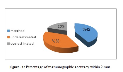
Eighty-four out of two hundred patients (42%) had the same tumour size
(± 2 mm) in both mammographic and histopathologic results. In 116/200 patients
there was difference in size. Furthermore, the tumour size was underestimated
in 76 (38%) patients, overestimated in 40 (20%) patients (Fig. 1).
The mean tumour size measured by
mammography and histopathology was 32.36±14.64 and 33.87±15.11 mm respectively.The mean value of
underestimation and overestimation of tumour size were 6.96 ± 4.70 and 5.3 ±
4.04 mm, respectively. Lastly, the
difference and correlation of the mean size between mammography and
histopathology were statistically significant t=-3.83, p=0.000 ;( r=0.93,
p<0.05). The mammography accurately determined the tumour
size (versus pathologic size) within 5 and 10mm, in 77 and 90% of cases, respectively
(Table I).
Table I: Distribution of actual accuracy to detect tumor size
Mammogram Vs histopathology within 2, 5 and 10 mm.
|
Tumor size
|
Accuracy within 2 mm
No. of patients (n=200)
%
|
Accuracy within 5 mm
No. of patients (n=200) %
|
Accuracy within 10 mm
No. of patients(n=200) %
|
|
Matched
|
84 42
|
154 77
|
180
90
|
|
Overestimated
|
76 38
|
12 6
|
4 2
|
|
Underestimated
|
40 20
|
34
17
|
16
8
|
The mean
number of dissected axillary lymph node was 20 (ranges: 10 – 43). Forty eight
patients (24%) had no lymph node metastasis, while 152 patients (76%) had lymph
node metastasis. The N0, N1, N2 and N3 status distribution was 24, 31, 25 and
20%, respectively. In axillary ultrasound, using the
lymph node morphology as a criteria for positivity (increase size, thick cortex
and loss of fatty hilum), sensitivity and specificity were found to be 87 and
67%, respectively. The positive predictive value (PPV) and the negative
predictive value (NPV) were 88 and 66% respectively. (Table II)
Table II:
Axillary
ultrasound (US) results.
|
%
|
No. of patients
(n=200)
|
Findings
|
|
63.5
|
127
|
True positive
|
|
18.5
|
37
|
True negative
|
|
8.5
|
17
|
False positive
|
|
9.5
|
19
|
False negative
|
Discussion
In breast carcinoma, tumour size and lymph node number are the two important prognostic factors. (24) In a study with 20-year follow-up, Rosen et al. reported a recurrence-free survival rate of 88% for <1.0 cm tumor, 72% for 1.1 to 3.0 cm tumours, and 59% for 3.1 to 5.0 cm tumours. (25) In a study by Hieken et al, mammography underestimated the tumour size in 60% of the patients, the mean underestimation of the breast tumor size was 3.5 ± 0.9 mm, for mammographically determined size (versus pathologic size) correlation, r, was 0.4, the mammogram accurately determined the tumor size within 2, 5, and 10 mm in 32, 65 and 85% of cases, respectively.(26).
In the present study 84/200 patients (42%) had the same tumor size (within 2 mm) in both mammography and histopathologic results. In 116/200 patient there was a difference in size. The mean value of difference estimated by mammography and histopathology was 1.51± 5.57 mm, while the minimum and maximum deference ranges from 1-20 mm. The tumour size was underestimated in 76 patients (38%), and it was overestimated in 40 patients (20%). Furthermore the mean value of underestimation and overestimation of tumour size were 6.96 ± 4.7 and 5.3 ± 4.04 mm, respectively. The mammography accurately determined the tumor size (versus pathologic size) within 5 and 10mm, in 77 and 90% of cases, respectively.
In the present study 84/200 patients (42%) had the same tumor size
(within 2 mm) in both mammography and histopathologic results. In 116/200
patient there was a difference in size.
The mean value of difference estimated by mammography and histopathology was
1.51± 5.57 mm, while the minimum and maximum deference ranges from 1-20 mm. The
tumour size was underestimated in 76 patients (38%), and it was overestimated
in 40 patients (20%). Furthermore the mean value of underestimation and
overestimation of tumour size were 6.96 ± 4.7
and 5.3 ± 4.04 mm, respectively. The mammography accurately determined the tumor size (versus pathologic size) within 5
and 10mm, in 77 and 90% of cases, respectively.
The total number of involved nodes gives a prognostic marker which is directly related to the recurrence rate and indirectly related to overall survival. In a study of 1,741 cases, the 10- year survival of patients with N0, N1, N2, and N3 was 75%, 62%, 42%, and 20% respectively.(27)
In a study done by Alvarez et al, on sonography of axilla without palpable nodes, if the size of the node (> 5 mm) or its visibility was used as a criterion for positivity, the sensitivity and specificity varied from 48.8 to 87.1% and from 55.6 to 97.3, respectively. On the other hand, if the
morphology of the node was used as the criterion for positivity, sensitivity and specificity varied from 26.4 to 75.9% and from 88.4 to 98.1%, respectively. If palpable and non-palpable nodes are included and if the size (> 5 mm) or visibility on sonography of the node was used as the criterion for positivity, sensitivity ranged from 66.1 to 72.7%, while specificity ranged from 44.1 to 97.9%.(28) Table III shows the sensitivity and the specificity of axillary ultrasonography in the detection of lymph node metastasis in ten international studies that used the lymph node size and the node morphology as criteria for positivity.
|
Table III:
Diagnostic accuracy of axillary sonography in patients with breast
carcinoma.
|
|
Study
|
Date
|
TP
|
TN
|
FP
|
FN
|
Sensitivity (%)
|
p
|
Specificity (%)
|
p
|
|
|
Size
criterion
|
|
|
|
|
|
|
|
|
|
|
|
Bruneton
et al. (29)
|
1986
|
16
|
37
|
1
|
6
|
72.7 (49.8–89.3)
|
|
97.4 (86.2–99.9)
|
|
|
|
Tate
et al. (30)
|
1989
|
39
|
61
|
20
|
20
|
66.1 (52.6–77.9)
|
|
75.3 (64.5–84.2)
|
|
|
|
Mustonen
et al. (31)
|
1990
|
12
|
46
|
1
|
6
|
66.7 (40.9–86.6)
|
|
97.9 (88.7–99.9)
|
|
|
|
Vaidya
et al. (32)
|
1996
|
78
|
78
|
9
|
35
|
69.0 (59.6–77.4)
|
|
89.7 (81.3–95.2)
|
|
|
|
Damera
et al. (33)
|
2003
|
46
|
45
|
57
|
18
|
71.8 (59.2–82.4)
|
|
44.1 (34.3–54.3)
|
|
|
|
Summaryb
|
|
|
|
|
|
68.4 (61.7–74.6)
|
|
87.7 (83.1–91.5)
|
|
|
|
Heterogeneityb,c
|
|
|
|
|
|
0.38
|
0.94
|
20.86
|
0.000
|
|
|
Summary
|
|
|
|
|
|
69.2 (63.4–74.6)
|
|
75.2 (70.4–79.6)
|
|
|
|
Heterogeneityc
|
|
|
|
|
|
0.67
|
0.95
|
90.27
|
0.000
|
|
|
Morphologic
criteriond
|
|
|
|
|
|
|
|
|
|
|
|
Lam
et al. (34)
|
1996
|
8
|
19
|
1
|
3
|
72.7 (39.0–94.0)
|
|
95.0 (75.1–99.8)
|
|
|
|
Yang
et al. (35)
|
1996
|
35
|
68
|
2
|
9
|
79.5 (64.7–90.2)
|
|
97.1 (90.0–99.6)
|
|
|
|
Verbanck
et al. (15)
|
1997
|
24
|
20
|
1
|
2
|
92.3 (74.9–99.1)
|
|
95.2 (76.2–99.9)
|
|
|
|
Yang
et al. (36)
|
1998
|
31
|
40
|
2
|
8
|
79.5 (63.5–90.7)
|
|
95.2 (83.8–99.4)
|
|
|
|
Sapino
et al. (37)
|
2003
|
60
|
144
|
35
|
28
|
68.2 (57.4–77.7)
|
|
80.4 (73.9–86.2)
|
|
|
|
Damera
et al. (33)
|
2003
|
35
|
83
|
19
|
29
|
54.7 (41.7–67.2)
|
|
81.4 (72.4–88.4)
|
|
|
|
Summaryb
|
|
|
|
|
|
81.7 (73.6–88.1)
|
|
96.1 (91.7–98.5)
|
|
|
|
Heterogeneityb,c
|
|
|
|
|
|
3.18
|
0.36
|
0.4
|
0.94
|
|
|
Summarye
|
|
|
|
|
|
71.0 (65.2–76.3)
|
|
86.2 (82.6–89.3)
|
|
|
|
Heterogeneityc,e
|
|
|
|
|
|
18.38
|
0. 003
|
23.53
|
0. 000
|
|
Note–Numbers
in parentheses are 95% confidence intervals. TP = true-positive, TN =
true-negative. FP = false-positive, FN = false-negative.
aStudies in which criterion for
classifying axillary node as positive was size
bIncludes
only studies in which gold standard was axillary lymph node dissection
c Chi-square
test, we used
dStudies in which criterion for
classifying axillary node as positive was morphologic or structural
eIncludes
studies in which gold standard was axillary lymph node dissection or sentinel
node biopsy
In our study we used the node size, thickening
of the cortex and loss of fatty hilum as a criterion for positivity. Therein, sensitivity and
specificity were 87 and 67%, respectively.
Based on the results of the current study we
believe that the inaccurate sizing of mammography can lead to unnecessary
mastectomies and increase the incidence of positive margin tumours, but even
this could happen, it will not affect the overall survival, and this actually
needs more data.
Conclusion
This study demonstrates that the mammography does not seem to be very accurate in
detecting the tumor size. Moreover, the axillary ultrasound is quite sensitive
and moderately specific in the diagnosis of axillary lymph node metastasis.
References
1. Khalil AM,
Ayad EE, El-Sheikh SA: Immunohistochemical expression of ckit in invasive
breast carcinoma of different nuclear grades. Med J Cairo Univ 2012;
80:345–351.
2. American Cancer Society.
Cancer Facts and Figures 2015. Atlanta, GA: American Cancer Society; 2015.
3. Jordanian
Ministry of Health, Jordan Cancer Registry, Cancer Incidence in Jordan 2012. Journal.2012; vol
(17):54-59.
4. Veronesi U, Galimberti V, Zurrida S,
Pigatto F, Veronesi P, Robertson C, et al. Sentinel lymph node biopsy as an
indicator for axillary dissection in early breast cancer. Eur J Cancer.
2001;37:454-8.
5. Cowen D, Jacquemier J, Houvenaeghel G,
Viens P, Puig B, Bardou VJ, et al. Local and
distant recurrence after conservative management of ‘very low-risk’ breast
cancer are dependent events: a 10-year follow-up. Int J Radiat Oncol Biol
Phys. 1998;41:801-807.
6. Dongen van JA,
Bartelink H, Fentiman IS, Lerut T, Mignolet F, Olthuis G, et al. Factors
influencing local relapse and survival and results of
salvage treatment after breast-conserving therapy in operable breast cancer: EORTC
trial 10801, breast conservation compared with mastectomy in TNM stage I and II
breast cancer. Eur J Cancer. 1992;28A(4-5):801-805.
7. Carter CL, Allen C, Henson DE.
Relation of tumor size, lymph node status, and survival in 24 740 breast cancer
cases. Cancer. 1989;63:181-187.
8. Sobin LH,
Wittekind C. TNM Classification of malignant tumours, Breast Tumours (ICD-O
C50).
7edn. Chichester, West Sussex; Hoboken: John Wiley & Sons; 2009.
9. Harirchi I, KarbakhshM , Kashefi A , Momtahen A J :
Breast Cancer in Iran: results of a
multi-center study, Asian Pacific J Cancer Prev. 2004;
5(1):24-27.
10.Cheung
YC, Wan YL,et al :Diagnostic performance of dual-energy contrast- enhanced subtracted mammography in
dense breasts compared to mammography alone: interobserver blind-reading analysis. Eur radiol. 2014;
24:2394-2403.
11.ChalaL,Endo E,Kim S,deCastro F Morae P,Cerri G et al:Gray‐scale sonography of solid breast masses: diagnosis of probably benign masses and reduction of the number of biopsies,J-Clin Ultrasound. 2007;35:9-19.
12. Lazarus E, Mainiero MB, Schepps B, Koelliker SL, Livingston LS BI-RADS Lexicon for
US and mammography: interobserver variability and positive predictive value, Radiology. 2006; 239: 385-391.
13.
Abdullah N, Mesurolle B, El-Khoury M, Kao E: Breast imaging reporting and
data system lexicon for US: interobserver agreement for assessment of breast masses.
Radiology. 2009;252: 665–672.
14. Houssami N, Ciatto S, Turner RM, Cody
HS, 3rd,
MacaskilP. Preoperative ultrasound-guided needle biopsy of axillary
nodes in invasive breast cancer: meta-analysis of its accuracy and utility in
staging the axilla. Ann Surg. 2011;254:243-251.
15. Verbanck J, Vandewiele I, De Winter
H, Tytgat J, Van Aelst F, Tanghe W. Value of axillary
ultrasonography and sonographically guided puncture of axillary nodes: a prospective study in 144
consecutive patients. J Clin Ultrasound. 1997;25:53-56.
16. Bonnema J, van Geel AN, van Ooijen B,
Mali SP, Tjiam SL, Henzen-Logmans SC, et al. Ultrasound-guided aspiration
biopsy for detection of nonpalpable axillary node metastases in breast cancer
patients: new diagnostic method. World J Surg.1997;21:270-274.
17. Deurloo EE, Tanis PJ, Gilhuijs
KG, Muller SH, Kröger R, Peterse JL, et al. Reduction in the number of
sentinel lymph node procedures by preoperative ultrasonography of the axilla in
breast cancer. Eur J Cancer. 2003;39:1068-1073.
18. Abe H, Schmidt RA,
Kulkarni K, Sennett CA, Mueller JS, Newstead GM. Axillary lymph nodes
suspicious for breast cancer metastasis: sampling with US-guided 14-gauge
core-needle biopsy--clinical experience in 100 patients. Radiology. 2009;250:41-49.
19. Britton PD, Goud A,
Godward S, Barter S, Freeman A, Gaskarth M, et al. Use of ultrasound-guided axillary
node core biopsy in staging of early breast cancer. Eur Radiol. 2009;19:561-569.
20. Podkrajsek M, Music MM,
Kadivec M, Zgajnar J, Besic N, Pogacnik A, et al. Role of ultrasound in the
preoperative staging of patients with breast cancer. Eur Radiol. 2005;15:1044-1050.
21. Koelliker SL, Chung MA,
Mainiero MB, Steinhoff MM, Cady B. Axillary lymph nodes:
US-guided fine-needle aspiration for initial staging of breast
cancer--correlation with primary tumor size. Radiology.2008;246:81-89
22. Duchesne N, Jaffey J,
Florack P, Duchesne S. Redefining ultrasound appearance criteria of positive
axillary lymph nodes. Can Assoc Radiol J. 2005;56:289-296.
23. Garcia-Ortega MJ, Benito
MA, Vahamonde EF, Torres PR, Velasco AB, Paredes MM. Pretreatment axillary
ultrasonography and core biopsy in patients with suspected breast cancer:
diagnostic accuracy and impact on management. Eur J Radiol. 2011;79:64-72.
24.
Greene FL, Page DI, Fleming ID. AJCC cancer staging manual, 6th
ed. New York: Springer-Verlag;2002.
25. Rosen EL, Blackwell KL, Baker JA, oo MS, Bentley RC, Yu D,
et al.
Accuracy of MRI in the detection of residual breast cancer after neoadjuvant
chemotherapy. AJR Am J Roentgenol. 2003; 181: 1275-1282.
26. Hieken
TJ1, Harrison J, Herreros J, Velasco JM : Correlating sonography, mammography,
and pathology in the assessment of breast cancer size. Am J Surg.
2001;182(4):351-354.
27. Fisher BJ, Perera FE, Cooke
AL,Opeitum A,Venkatesan V,Rashid Dar A,et al. Long-term follow-up of axillary
node-positive breast cancer patients receiving adjuvant systemic therapy alone:
patterns of recurrence. Int J Radiat Oncol Biol Phys. 1997; 38: 541-550.
28.
Alvarez S, Anorbe E, Alcorta P, Lopez F, Alonso I, Cortes J.Role of sonography in the
diagnosis of axillary lymph node metastases in breast cancer: a systematic
review. AJR Am J Roentgenology. 2006;186:1342-1348.
29.Bruneton JN, Caramella E, Héry M, Aubanel D, Manzino
JJ, Picard JL. Axillary lymph node metastases in breast cancer:
preoperative detection with US. Radiology.1986; 158:325-326.
30.
Tate JJ, Lewis V, Archer T, Guyer PG, Royle GT, Taylor I. Ultrasound detection of axillary lymph node metastases in breast
cancer. Eur J Surg Oncol. 1989; 15:139-141.
31.Mustonen P, Farin P,
Kosunen O. Ultrasonographic detection of metastatic axillary
lymph nodes in breast cancer. Ann Chir Gynaecol. 1990; 79:15-18.
32.
Vaidya JS, Vyas JJ, Thakur MH, Khandelwal KC, Mittra I. Role of ultrasonography to detect axillary node involvement in
operable breast cancer. Eur J Surg Oncol.1996; 22:140-143.
33. Damera A, Evans AJ, Cornford EJ, Wilson ARM,
Burrell HD, James JJ,et al. Diagnosis of
axillary nodal metastases by ultrasound-guided core biopsy in primary operable
breast cancer. Br J Cancer 2003; 89:1310-1313.
34. Lam WW, Yang WT,
Chan YL, Stewart IE, Metreweli C, King W. Detection
of axillary lymph node metastases in breast carcinoma by technetium-99m
sestamibi breast scintigraphy, ultrasound and conventional mammography. Eur
J Nucl Med. 1996; 23:498-503.
35. Yang WT, Ahuja A,
Tang A, Suen M, King W, Metreweli C. High
resolution sonographic detection of axillary lymph node metastases in breast
cancer. J Ultrasound Med 1996; 16:241-246; erratum in: J Ultrasound Med.
1996; 15:644.
36.
Yang WT, Metreweli C. Colour Doppler flow in normal axillary lymph nodes. Br
J Radiol. 1998; 71:381-383.
37.
Sapino A, Cassoni P, Zanon E, Fraire F,
Croce S, Coluccia C, et al. Ultrasonographically-guided
fine-needle aspiration of axillary lymph nodes: role in breast cancer
management. Br J Cancer. 2003; 88:702-706.